- Case-Based Roundtable
- General Dermatology
- Eczema
- Chronic Hand Eczema
- Alopecia
- Aesthetics
- Vitiligo
- COVID-19
- Actinic Keratosis
- Precision Medicine and Biologics
- Rare Disease
- Wound Care
- Rosacea
- Psoriasis
- Psoriatic Arthritis
- Atopic Dermatitis
- Melasma
- NP and PA
- Skin Cancer
- Hidradenitis Suppurativa
- Drug Watch
- Pigmentary Disorders
- Acne
- Pediatric Dermatology
- Practice Management
- Prurigo Nodularis
- Buy-and-Bill
Publication
Article
Dermatology Times
AAD updates cutaneous melanoma guidelines
Author(s):
Treatment options for metastatic melanoma have greatly improved, including in the adjuvant setting, and these advances are reflected in the recently updated American Academy of Dermatology clinical guidelines on the management of primary cutaneous melanoma. Find out what has changed in this article.
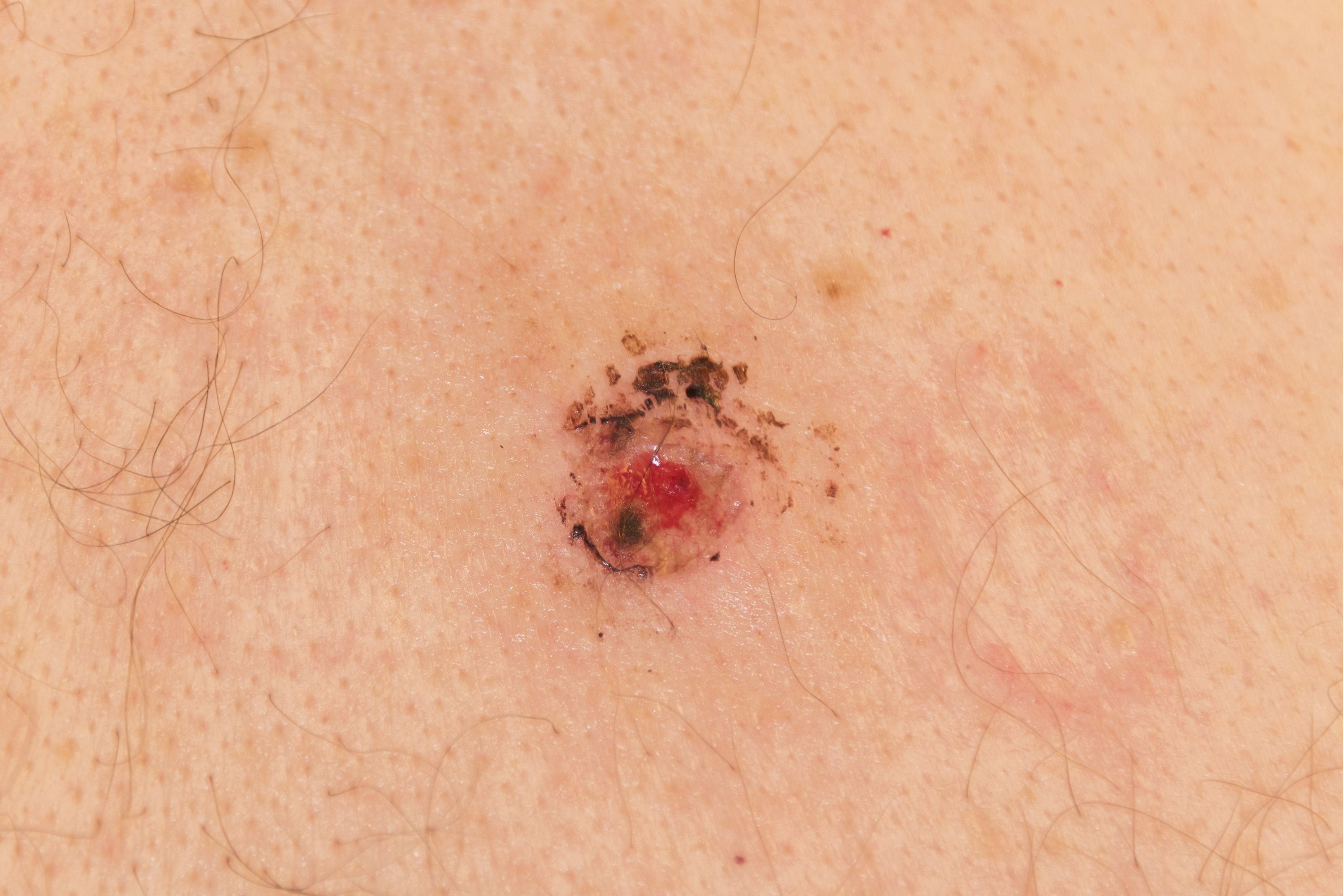
The American Academy of Dermatology recently updated its clinical guidelines on the management of primary cutaneous melanoma. Find out what has changed in this article. (JimVallee - stock.adobe.com).

Dr. Susan Swetter

Treatment options for metastatic melanoma have greatly improved, including in the adjuvant setting, and these advances are reflected in the recently updated American Academy of Dermatology clinical guidelines on the management of primary cutaneous melanoma.
The guidelines, last revised in 2011, also take into account the American Joint Committee on Cancer (AJCC) latest recommendations on staging and the use of information from biopsy, pathology, primary surgery, and sentinel lymph node biopsy (SNLB).
“It is only in the past year that effective systemic adjuvant therapies for surgically resected stage III melanoma have been FDA-approved,” says Susan Swetter, M.D., chair of the guidelines working group and director of the Pigmented Lesion and Melanoma Program at Stanford University Medical Center and Cancer Institute.
“Adjuvant anti-PD-1 monotherapy and combination BRAF/MEK inhibitors are now associated with improved disease-free and overall survival, making accurate staging of patients with cutaneous melanoma even more important. This currently involves pathological staging with sentinel lymph node biopsy in appropriate patients,” she said.
BIOPSY
Although various molecular and imaging techniques have been studied as adjuncts to histopathologic assessment, biopsy remains the first step to establish a definitive diagnosis of cutaneous melanoma (CM) as it enables tumors to be categorized according to their thickness and appearance.
The recommended technique is a narrow excisional/complete biopsy with 1- to 3-mm margins that encompass the entire breadth of lesion and is of sufficient depth to prevent transection at the base. It can be a fusiform/elliptical or punch excision or deep shave/saucerization removal to a depth below the anticipated plane of the lesion.
Partial/incomplete sampling (incisional biopsy) is acceptable when the location of the lesion requires, such as when it is facial or acral. A narrow-margin excisional biopsy may then be performed if required for diagnosis or microstaging, but this is not usually necessary if the initial specimen meets the criteria for sentinel lymph node biopsy.
STAGING
Under the AJCC 8th edition staging for cutaneous melanoma for T1 melanoma-which have a thickness of ≤1 mm-0.8 mm thickness is now the threshold for T1a (classified as <0.8 mm without ulceration), whereas T1b is defined as 0.8 – 1.0 mm thickness with or without ulceration or <0.8 mm thickness with ulceration.
Melanoma is classified T2 if thickness is >1.0 to 2.0 mm, T3 at >2.0 to 4.0 mm and T4 at >4.0 mm, and for all these thicknesses ‘a’ indicates no ulceration and ‘b’ indicates ulceration.
In addition, N and M classification under AJCC staging specifies the quantity and extent of node involvement, and location of metastatic spread respectively.
PATHOLOGY
When a biopsy of a suspicious lesion is performed, the guideline says that the pathologist should be given essential information about the patient including their age, sex and precise anatomic location of the biopsy site.
It is strongly recommended that the clinician include their clinical impression and differential diagnosis, the size of the lesion, and intent of the biopsy (ie, excisional vs partial, and type of diagnostic biopsy performed (elliptical/fusiform, deep shave/saucerization, broad shave, or punch).
Macroscopic satellites around the clinical lesion should be pointed out, as they escalate cutaneous melanoma to stage III and can be associated with microscopic satellites in the primary tumor, and any clinical photographs of the site provided for review.
Other useful information that may be useful for the pathologist include the level of suspicion for cutaneous melanoma, clinical description and history of the lesion beyond size (including whether there has been a change in the lesion or previous biopsy), and dermoscopic features (with or without an accompanying photograph).
SURGICAL MANAGEMENT
Surgery remains the first line treatment for cutaneous melanoma of any thickness as well as melanoma in situ (melanoma cells in epidermis; stage 0), so following initial biopsy, wider and deeper excision is performed to ensure complete removal of the lesion, confirm histologically clear margins, and reduce the risk of local recurrence.
Surgical margins should be based on tumor thickness and greater for thicker tumors. For invasive cutaneous melanoma (CM) they should be >1 cm and <2 cm (1 cm is recommended for 1mm tumours and 2cm for 2mm tumours) and measured clinically around the primary tumor, although they may be narrower to accommodate function and/or anatomic location. For melanoma in situ, wide excision with 0.5- to 1.0-cm margins is recommended.
The recommended depth of excision is to (but not including) the fascia.
Recommended surgical excision margins are measured from the edge of the lesion or prior biopsy at the time of surgery and are not histologic margins as measured by the pathologist.
Sentinel lymph node biopsy, when indicated, should be performed before wide excision of the primary tumor, and in the same operative setting, whenever possible.
SENTINEL NODE BIOPSY
Sentinel node biopsy is not recommended for patients with melanoma in situ or for most T1a CM (<0.8 mm without ulceration).
The procedure should be discussed with and offered to appropriate patients with CM>1 mm thickness (>T2a), including T4. It should be also be discussed and considered in patients with T1b CM (<0.8 mm with ulceration or 0.8-1.0 mm with or without ulceration), although rates of SLN positivity are relatively low in this group.
The procedure may be considered for T1a cutaneous melanoma if other adverse features are present, including young patient age, presence of lymphovascular invasion, deep biopsy margin (if close to 0.8 mm) or high mitotic rate.
MOHS, IMIQUIMOD & RADIATION THERAPY
There has been much progress in the use of Mohs micrographic surgery and other staged surgery techniques (utilizing permanent sections) for treatment of melanoma in situ on anatomically constrained sites (typically the lentigo maligna subtype).
However, Dr. Swetter says: “Current data are insufficient for the guidelines to recommend Mohs surgery for invasive cutaneous melanoma, in which the use of surgical margins less than 1 cm has not been adequately studied worldwide.
“Non-surgical approaches, including the off-label use of imiquimod and traditional forms of radiation therapy, may be considered if surgery is impractical or contraindicated, and only for melanoma in situ, lentigo maligna type, as cure rates are lower. These modalities all require further prospective-investigation, which is in progress in the US and abroad.”
SURVEILLANCE
Although the optimal interval and duration of follow-up for CM are not well defined, the guideline working group recommends that patients with CM be monitored regularly following diagnosis, particularly for tumors at increased risk of recurrence (ie, >2.0 mm in thickness and/or with ulceration, lymphovascular invasion, high mitotic rate).
Although most metastases occur in the first 1 to 3 years after treatment of the primary tumor, skin examinations for life are generally recommended.
For patients with melanoma in situ (stage 0), reviews are recommended at least every 6 to 12 months for 1 to 2 years and then annually. Review should involve a physical examination to check for local recurrence and a full skin check to spot any new primary cutaneous melanoma.
Patients with stage IA-IIA should also be reviewed every 6 to 12 months initially, but for 2-5 years, and then annually. These reviews should involve a comprehensive history and physical examination focused on the skin and regional lymph nodes. Patients with stage IIB and greater tumours should receive a similar review every 3-6 months for the first 2 years, then at least every 6 months for the next 3-5 years and then at least annually. Patients with stage IIB and greater tumours also likely to benefit from radiological testing for 3 two five years.
KEY QUESTIONS
The guidelines working group considered the key clinical questions that frequently arise in the management of cutaneous melanoma and three new topics emerged: Melanoma in pregnancy, genetic testing for hereditary melanoma, and dermatologic toxicities associated with newer melanoma drugs (eg targeted agents such as BRAF/MEK inhibitors and immune checkpoint inhibitors) which dermatologists will see more of given their broader use across the cancer spectrum.
The guidelines say that a diagnosis of cutaneous melanoma during pregnancy does not alter prognosis or outcome for the woman, however, work-up and treatment must take the safety of the fetus into consideration. The approach taken to melanocytic nevi in the pregnant woman should be identical to that in the nonpregnant patient. Any changing nevus during pregnancy should be evaluated and subjected to biopsy if clinically and/or dermoscopically concerning.
Dermatologists should collaborate with oncologists for management of cutaneous toxicity during BRAF/MEK kinase or immune checkpoint inhibitor therapy, the guidelines say, because appropriate recognition and control of skin side effects may improve the quality of life of patients with cutaneous melanoma and avoid unnecessary interruption of medication. The frequency of dermatologic assessment depends on the agent(s) being used, age of the patient, underlying skin cancer risk factors and/or potential role of skin findings as a biomarker for response.
Assessment is recommended every 2-4 weeks for the first 3 months of BRAF inhibitor monotherapy for patients with numerous squamoproliferative neoplasms, although combination BRAF/MEK inhibition is standard and associated with less skin toxicity. With immune checkpoint inhibitors, it should take place within the first month of therapy and continue as needed. Patients with atopic dermatitis, psoriasis, or other autoimmune dermatoses, should be seen by a dermatologist for pre-emptive counseling and treatment before initiation of therapy.
THE FUTURE
Novel molecular techniques are being studied for melanoma prognostication, to help determine which patients should be treated and followed more intensively.
“These techniques require ongoing prospective validation studies to determine their predictive value, how well they compare with and add to known histopathologic factors (and prognostic prediction models based on AJCC 8th edition staging), and how best to incorporate them into clinical practice. This is an exciting area that dermatologist can look forward to in the future,” Dr. Swetter says.
“As molecular techniques are further investigated, we anticipate improvement in our ability to determine which patients benefit most from surgery and/or systemic therapy. As such, the guidelines emphasize the advantages of multidisciplinary collaboration among dermatologists, surgeons, and medical oncologists, particularly for those patients at higher risk of disease recurrence,” she said. Â
References
Swetter SM et al. “Guidelines of care for the management of primary cutaneous melanoma,” Journal of the American Academy of Dermatology, Oct. 27, 2018. https://doi.org/10.1016/j.jaad.2018.08.055
Bichakjian CK, Halpern AC, Johnson TM, et al. “Guidelines of care for the management of primary cutaneous melanoma.” American Academy of Dermatology. J Am Acad Dermatol. 2011;65:1032-1047.
Gershenwald JE, Scolyer RA, Hess KR, et al. “Melanoma of the skin.” In: Amin MB, Edge SB, Greene FL, et al. eds. AJCC Cancer Staging Manual. 8th ed. New York, NY: Springer International Publishing; 2017.


