- Case-Based Roundtable
- General Dermatology
- Eczema
- Chronic Hand Eczema
- Alopecia
- Aesthetics
- Vitiligo
- COVID-19
- Actinic Keratosis
- Precision Medicine and Biologics
- Rare Disease
- Wound Care
- Rosacea
- Psoriasis
- Psoriatic Arthritis
- Atopic Dermatitis
- Melasma
- NP and PA
- Skin Cancer
- Hidradenitis Suppurativa
- Drug Watch
- Pigmentary Disorders
- Acne
- Pediatric Dermatology
- Practice Management
- Prurigo Nodularis
- Buy-and-Bill
Publication
Article
Dermatology Times
Master the Clues that Mask Malignancy
Author(s):
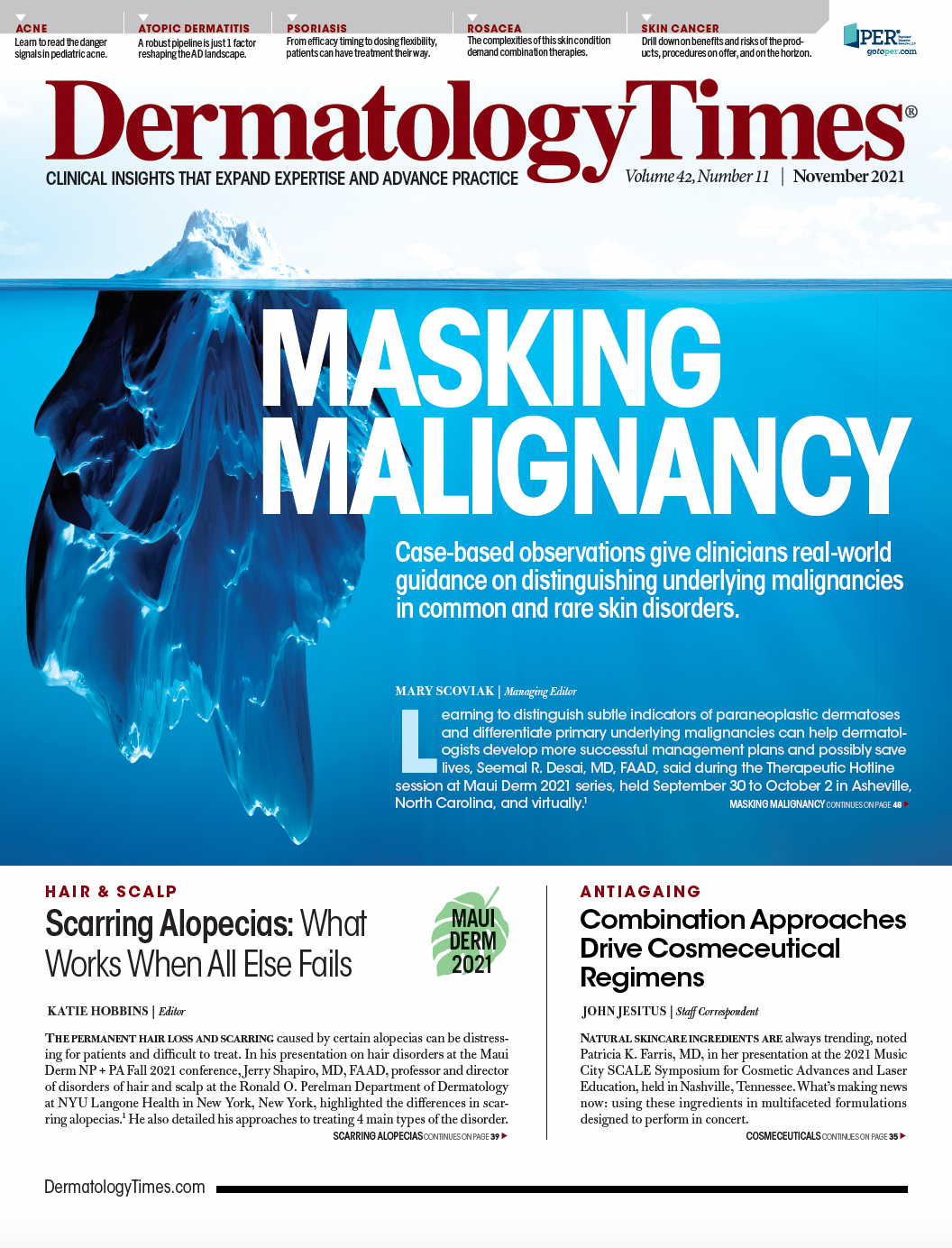
Case-based observations offered clinicians real-world guidance on distinguishing underlying malignancies in the Therapeutic Hotline session at Maui Derm NP+PA Fall 2021.
Learning to distinguish subtle indicators that point to paraneoplastic dermatoses and differentiate primary underlying malignancies in various disorders can help dermatologists develop more successful management plans and possibly save lives, Seemal Desai, MD, FAAD, said in the Therapeutic Hotline session at Maui Derm NP+PA Fall 2021, held September 30 to October 2, 2021, live in Asheville, North Carolina, as well as virtually.1
For his case-based presentation, Desai focused on assessing key factors that would raise suspicions about the presence of a malignant condition.
“Never say never,” said Desai, who is a clinical assistant professor in the Department of Dermatology at the University of Texas Southwestern Medical Center in Dallas, Texas. “Always consider an underlying malignancy when dealing with an atypical presentation of well-documented disorder.”
He offered these strategies for detecting and treating conditions that could negatively impact the patient's daily or become life-threatening.
Papulosquamous Disorders: Acanthosis Nigricans; acquired ichthyosis; tripe palms; Leser-Trélat syndrome and acrokeratosis paraneoplastica (Bazex syndrome).
In one case, a 73-year-old white male presented with scaly lesions on his hands, elbows, knees and abdomen for 6 months. His condition was non-pruritic. He had been experiencing severe weight loss for over 6 months,and was having difficulty swallowing. The combination of lesions and gastric suggested a diagnosis of malignant Acanthosis Nigricans, said Desai.
“Associated malignancies included gastric, esophageal, ovarian, endometrial, gallbladder, lung, bladder and others,” Desai said. “Nearly all are adenocarcinomas, of which 70% to 90% are intra-abdominal and 50% to 60% are gastric. The cancer is usually well-established when cutaneous signs appear and may improve with chemotherapy with 5 fluorouracil, cisplatin and epirubicin.”2
Tripe Palms
Assessing the possibility of malignacies underlying tripe palms underscores the need to consider all factors that could impact the diagnosis, according to Desai.
He pointed out that this condition is associated with lung carcinoma in more than 90% of cases. However, when patients present with both tripe palms and Acanthosis Nigricans, the case suggests an adenocarcinoma, typically gastric, according to Desai. He noted that 60% of tripe palms present before or around the same time as the diagnosis of the tumor and Based on current research, the condition does not have a race or gender predisposition, he added.3
Interface Dermatitides: Paraneoplastic dermatomyositis and paraneoplastic pemphigus.
“Dermatomyositis is a very common disease, but a scary one,” Desai said. “The malignancy associated with this condition usually is seen in patients in their 5th or 6th decade. Skin findings often precede the development of malignancy by a year.”
Female patients should be scanned for ovarian because dermatomyositis is highly linked to ovarian cancer, he added. He advised recommending gynecological exams every 3 to 6 months and having other pertinent health screenings. “Urge your female patients to see their OB/GYN or primary physician,” he said. “Talk to them. Take their history. Are their pants tighter? Is there unusual menstrual bleeding? Does their stomach feel full? If so, look for ovarian tumors.”
According to Desai, “treatment of the underlying malignancy usually clears the skin changes.”
Reactive Erythemas and Neutrophilic Dermatoses: Erythema gyratum repens; necrolytic migratory erythema; Sweet’s syndrome, and pyoderma gangrenosum.
Desai used the example of a patient with Sweet’s syndrome to illustrate the challenges of distinguishing malignancies. He pointed out that hematologic disorders such as Acute Myeloblastic Leukemia make up 85% of associated malignancies with genitourinary tumors, the most common solid tumors associated with Sweet’s syndrome. “Sweet’s should be treated as hematologic until proven otherwise, and patients should be receiving specific screenings including blood counts, peripheral smear, screening for GU malignancies, and repeat biopsies as needed,” he said.
Crohn’s Disease and Ulcerative Colitis
Fistulas are the first sign of Crohn’s Disease and can often be seen after procedures Metastatic Crohn’s disease can go to the skin. Skin lesions appear in 10-30% of Crohn’s cases. Crohn’s can involve lymph nodes and be associated with systemic sequelae. Rare metastatic Crohn’s can cause lesions that mimic erythema nodulosum.
“One of the key things to avoid with patients with Crohn’s who have fistulas is them going to a surgeon to have the fistula surgically treated,” Desai said. “That’s just going to make it worse.”
Patients with ulcerative colitis typically present with bloody diarrhea. UC has numerous skin manifestations and is usually linked to underlying bowel disease or other pathologies but can be associated with serious complications such as colorectal cancer. Erythema nodosum, pyoderma gangrenosum, apthous stomatitis, aseptic pustular eruptions and, rarely, pyoderma vegetans are all associated with UC.
Whatever the condition, Desai advised thinking outside the box to investigate hard-to-identify malignancies. “Full body skin scans are critical. Get patients in a gown and look at them head to toe,” he said. “Get a tissue culture. Chest x-rays are easy and affordable ways to investigate suspicious factors in the lungs. Do biopsies, but also consider tests beyond those typically used in dermatology. Take time to talk with the primary physicians or other specialists on the patient’s care team and let them know you’re part of that team.”
In addition, he recommended using recent data from the literature to enable clinicians to differentiate primary underlying malignancies and their most common type of skin presentation to develop treatment and workup plans for patients presenting with skin conditions suspicious for underlying malignancy and systemic disease.
“For all of these cases, realize that these patients are in our offices,” Desai said. “Some of these cases are rare, but rare things are going to happen. We have the opportunity to change, transform, and maybe save their lives.”
Disclosure:
Desai reported no relevant conflicts of interest in relation to this presentation.
References:
- Desai S, High W. Therapeutic Hotline. Presented at: Maui Derm NP+PA Fall 2021; September 30 to October 2, 2021; in Asheville, North Carolina, and virtual.
- Longshore SJ, Taylor JS, Kennedy A, Nurko S. Malignant acanthosis nigricans and endometrioid adenocarcinoma of the parametrium: the search for malignancy. J Am Acad Dermatol. 2003;49(3):541-543. doi:10.1067/s0190-9622(03)00913-7
- Cohen PR, Grossman ME, Almeida L, Kurzrock R. Tripe palms and malignancy. J Clin Oncol. 1989;7(5):669-678. doi:10.1200/JCO.1989.7.5.669


