- Case-Based Roundtable
- General Dermatology
- Eczema
- Chronic Hand Eczema
- Alopecia
- Aesthetics
- Vitiligo
- COVID-19
- Actinic Keratosis
- Precision Medicine and Biologics
- Rare Disease
- Wound Care
- Rosacea
- Psoriasis
- Psoriatic Arthritis
- Atopic Dermatitis
- Melasma
- NP and PA
- Skin Cancer
- Hidradenitis Suppurativa
- Drug Watch
- Pigmentary Disorders
- Acne
- Pediatric Dermatology
- Practice Management
- Prurigo Nodularis
- Buy-and-Bill
News
Article
QUIZ RECAP: Advanced Skin Cancer Awareness
Author(s):
Earlier this week, we shared our second Skin Cancer Awareness Month quiz. Review the answers and your responses below.
This week we asked the question: How much do you know about advanced skin cancer awareness?
Haven't taken our quiz yet? Pause before reading below and follow this link to complete it: here.
Below, we recap our second quiz and the correct answers to each question.
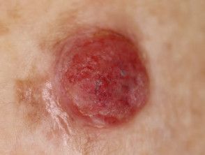
Question 1: What are the clinical features of amelanotic melanoma, and why is it often challenging to diagnose?
Response options:
- Presence of pigmented network, easily diagnosable
- Pink or flesh-colored lesion, challenging due to lack of pigment
- Regular borders, often misdiagnosed as benign
- Rapidly changing lesion, easily recognizable
Correct response option: Pink or flesh-colored lesion, challenging due to lack of pigment
Amelanotic melanoma, characterized by a lack of pigment, presents diagnostic challenges. History of lesion changes is crucial. Examining the whole skin surface is vital as other pigmented lesions may offer clues. Dermoscopy, assessing vessel morphology especially in unpigmented lesions, aids diagnosis.1
Question 2: What is the significance of the histopathological finding "lentiginous growth pattern" in the diagnosis of melanoma?
Response options:
- Indicates a benign lesion
- Suggests radial growth phase melanoma
- Indicates metastasis
- Indicates ulceration
Correct response option: Suggests radial growth phase melanoma
"Lentiginous" describes the radial growth phase of a tumor before it invades the dermis. This term may not always be paired with "acral," potentially leading to confusion among pathologists and clinicians.2
Question 3: How does dermatofibrosarcoma protuberans (DFSP) clinically differ from other cutaneous malignancies, and what is its characteristic histopathological feature?
Response options:
- Presents as a rapidly growing nodule, with no characteristic features
- Presents as a slowly growing, indurated plaque or nodule, with a storiform pattern histopathologically
- Presents as a blue-black nodule, with a characteristic "cartwheel" pattern histopathologically
- Presents as a pearly papule, with no characteristic histopathological features
Correct response option: Presents as a slowly growing, indurated plaque or nodule, with a storiform pattern histopathologically
DFSP typically arises on the trunk or proximal extremities, presenting as an indurated plaque. It is a low-grade malignancy, slow-growing, and often asymptomatic. Initially, it manifests as a firm papule or plaque, evolving over months or years into a nodule.3
Question 4: What are the key clinical and histological differences between lentigo maligna and lentigo maligna melanoma?
Response options:
- Lentigo maligna presents as a nodule, while lentigo maligna melanoma presents as a large, irregularly pigmented macule
- Lentigo maligna shows invasion beyond the epidermis histologically, while lentigo maligna melanoma does not
- Lentigo maligna melanoma presents with a regular border, while lentigo maligna has irregular borders
- Lentigo maligna melanoma typically regresses spontaneously, while lentigo maligna does not
Correct response option: Lentigo maligna presents as a nodule, while lentigo maligna melanoma presents as a large, irregularly pigmented macule
Lentigo maligna (LM) typically appears as an irregular brown macule on sun-damaged skin, especially on the head and neck of elderly individuals. First described as "Hutchinson’s melanotic freckle," it was once considered benign or precancerous. Today, LM is defined as melanoma in situ, confined to the epidermis. If invasive, it is termed lentigo maligna melanoma.4
Question 5: True or False: Merkel cell carcinoma (MCC) can mimic other benign skin lesions, making it challenging to diagnose clinically.
Response options:
- True
- False
- Partially true
- True only in elderly patients
Correct response option: True
Due to its rarity and similarity to other skin conditions, diagnosing Merkel cell carcinoma can be challenging, often mistaken for benign tumors or cysts. By the time it is correctly identified, it may have spread to lymph nodes. Early and accurate diagnosis requires specific tests and experienced health care providers.5
References
- Al-Ani A. Amelanotic melanoma. DermNet. January 2018. Accessed May 16, 2024. https://dermnetnz.org/topics/amelanotic-melanoma#:~:text=amelanotic%20melanoma%20diagnosed%3F-,Clinical%20features,considered%20in%20the%20differential%20diagnosis
- Basurto-Lozada P, Molina-Aguilar C, Castaneda-Garcia C, et al. Acral lentiginous melanoma: Basic facts, biological characteristics and research perspectives of an understudied disease. Pigment Cell Melanoma Res. 2021;34(1):59-71. doi:10.1111/pcmr.12885
- Kim MJ, Hur MS, Choi BG, et al. Pedunculated nodules as a variant of dermatofibrosarcoma protuberans. Ann Dermatol. 2016;28(5):629-631. doi:10.5021/ad.2016.28.5.629
- Xiong M, Charifa A, Chen CSJ. Lentigo maligna melanoma. StatPearls. Updated October 31, 2022. Accessed May 16, 2024. https://www.ncbi.nlm.nih.gov/books/NBK482163/
- Merkel cell carcinoma. Cleveland Clinic. Accessed May 16, 2024. https://my.clevelandclinic.org/services/merkel-cell-carcinoma-treatment#:~:text=MCC%20growths%20can%20be%20firm,you%20the%20care%20you%20need


