- Case-Based Roundtable
- General Dermatology
- Eczema
- Chronic Hand Eczema
- Alopecia
- Aesthetics
- Vitiligo
- COVID-19
- Actinic Keratosis
- Precision Medicine and Biologics
- Rare Disease
- Wound Care
- Rosacea
- Psoriasis
- Psoriatic Arthritis
- Atopic Dermatitis
- Melasma
- NP and PA
- Skin Cancer
- Hidradenitis Suppurativa
- Drug Watch
- Pigmentary Disorders
- Acne
- Pediatric Dermatology
- Practice Management
- Prurigo Nodularis
- Buy-and-Bill
Video
Presentation and Diagnosis of Atopic Dermatitis in Pediatrics
Author(s):
Brittany Craiglow, MD; Raj Chovatiya, MD, PhD; and Joshua Zeichner, MD, discuss the presentation of pediatric patients with atopic dermatitis and how to properly assess and diagnose pediatric patients’ atopic dermatitis.
Joshua Zeichner, MD: Let’s talk about the presentation of atopic dermatitis [AD]. It presents a little differently depending on the age of the patients. Brit, tell us a little about this.
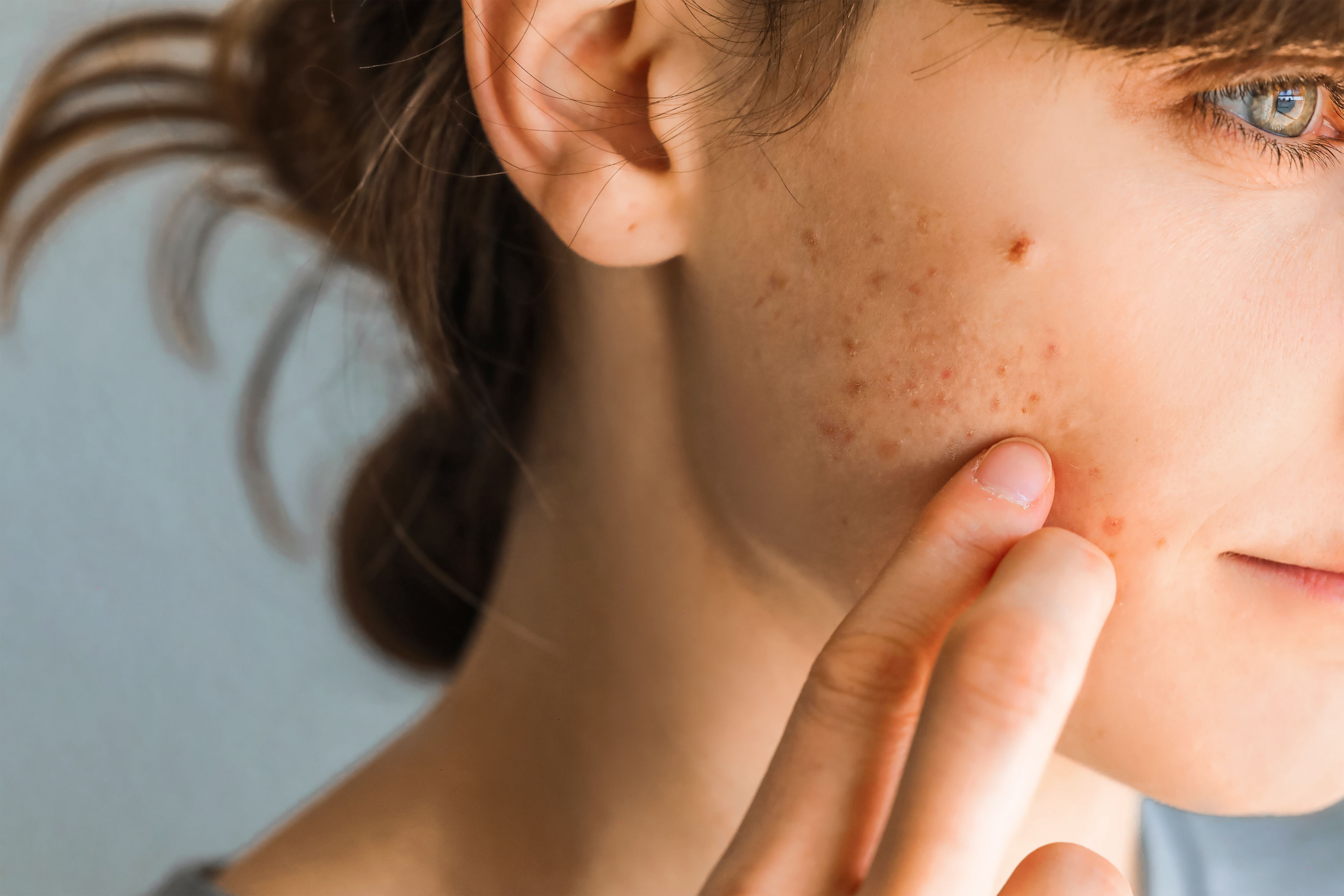
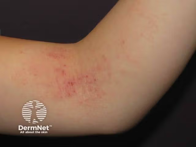
Brittany Craiglow, MD: Traditionally, we learned that atopic dermatitis is red, oozy, scaly, and dry—and a lot of times it is—but it’s important to understand that it can look very different in different people. Classically in infants, it’s going to be more extensor surface–heavy on the face, those stoplight red cheeks that you get. Then as kids get a little older and start to walk, it switches to more of that adult presentation, where you have the antecubital fossa and popliteal fossa heavily involved. Many patients are going to be very red, dry, and crusty. But especially in patients with darker skin types, it’s important to note that it doesn’t always look like that. Sometimes from the door, you might not even be able to recognize it. But when you get up close, you’ll see that the patient might be covered in follicular-based papules or have more of a prurigo nodularis type of presentation.
Another important point is that patients with AD have inflammation, even when you don’t necessarily see anything on their skin. Sometimes parents will say, “Her skin looks pretty good, but she’s still itchy all the time.” Our assessment of the patient isn’t about only what we see. It’s about their experience with the disease. Every so often, residents will tell me, “The patient is here for atopic dermatitis. It doesn’t look that bad.” And I say, “It doesn’t look that bad to you? Or it doesn’t look that bad to them?” We have to remind ourselves that we see this spectrum. We see a patient who’s erythrodermic, but that doesn’t mean that the patient who just has the antecubes and popliteal fossae isn’t having a big impact on their quality of life. It can look different in different people. Understanding the patient experience and the itch is huge. That can help drive our treatment more so than what we see.
Joshua Zeichner, MD: I’m so glad that you brought up those 2 points. I want to highlight that a little more. No. 1 is skin of color. For so long, we’ve seen photos of atopic dermatitis in White patients, and we need to recognize the different clinical presentations that we see in skin of color. Whether we’re talking about African American patients, Asian patients, or South Asian patients, we need to recognize that not all atopic dermatitis looks the same.
The other point that I love that you brought up is patient experience. When we talk to our patients, we can’t make assumptions about how much it impacts their quality of life based on what we’re seeing. You always need to have open-ended conversations with patients, asking them, “Was today a good day? Is today a bad day or an average day? How is this affecting you?” You can’t quantify itch based on the severity of the lesions that you’re seeing on the skin.
Brittany Craiglow, MD: We have to remind ourselves that we sometimes catch them on a good day, but even on a good day, many patients are worried about their next flare. The chronicity of the disease and the ebb and flow is difficult physically and psychologically. Finding out where they’ve been over the last several months since we’ve seen them is more important than where they are at that moment in time.
Joshua Zeichner, MD: This bridges nicely to my next question, which is the way that you guys clinically assess your patients with atopic dermatitis and make the diagnosis. Raj, let me turn this over to you.
Raj Chovatiya, MD, PhD: I’m happy to wax poetic on this one. The most important thing to think about is the heterogeneity of disease. That’s one of the things I love talking about regarding atopic dermatitis. The burden and presentation varies so much from person to person that we’ve tried to boil it down to anchoring on 1 specific thing we’re looking for when it comes to making assessments of severity. But in reality, there are a lot of things going into this calculation.
To take a step back, in terms of diagnosis, a lot of times people come to the clinic, and your gut tells you based on the story and what you’re seeing that this is probably atopic dermatitis. But atopic dermatitis is officially a clinically diagnosed disease. There are a couple of different systems for diagnosis. There isn’t 1 specific system that’s used quite a bit, but the first codified one was the Hanifin and Rajka criteria. Typically, you’re using a series of major and minor criteria.
In practice, most people aren’t doing this, but for the audience today, typically what you’re looking for is pruritus and some type of typical morphology or distribution. This includes lichenification or linearity in adults, facial extensor involvement in infants and children, typically a chronic relapsing dermatitis, and a personal family history of acne. Then there’s a number of minor criteria you can go for. People have tried to shorten this up over years and make easier, more clinically relevant criteria. A common one you’ll see a lot in the literature is the UK Working Party’s Diagnostic Criteria for Atopic Dermatitis. This is where people are saying someone has to have an itchy skin condition and then a couple of other small factors: things like flexure involvement, a history of atopy, and generalized dry skin.
From a practical standpoint, the AAD, the American Academy of Dermatology, largely recommends that you’re talking about some type of dermatitis condition and chronic itch, and then you put the pieces together. There are a lot of ways to look at it. But once you’ve made the diagnosis, when it comes to figuring out, “Is this mild? Is this moderate? Is this severe?” Oftentimes we’re anchoring on the extent. What’s the body surface area? But that’s just 1 aspect of disease that doesn’t perfectly correlate.
There are measures of lesion severity. Oftentimes people use gestalt or Investigator’s Global Assessments to rank someone as mild, moderate, or severe, but that’s usually looking at a very small defined area. People forget that there are lots of different morphologies, topographic distributions, comorbid burdens, and quality-of-life effects. There are different responses to previous medications. There are huge differences in longitudinal course when it relates to the disease. I like to think about all of those in context together when I’m trying to assess severity rather than boiling it down to only 1 specific aspect.
Joshua Zeichner, MD: Are there any pearls that you have on specific ways atopic dermatitis presents in different skin types?
Raj Chovatiya, MD, PhD: That’s a huge one that Brittany very nicely started bringing up in our previous discussion. I’m happy to continue along. One big question people always ask is: “If you aren’t used to looking at different types of skin pigmentation and if erythema is such an important component of what you’re looking for, how do you best measure severity using the tools we have?” Redness can look like a variety of things. It can look brown, purplish, like hyperpigmentation, violaceous—you name it. They’re all ways to get at inflammation. That’s 1 challenge that can occur from skin type to skin type.
The other thing I’ll bring up is that there are different types of lesions that all fall under the atopic dermatitis umbrella. If you were to ask someone about atopic dermatitis, they’d say, “Inside the elbows, back of the knees, flexural areas, done deal.” But if you look at a nice systematic review and meta-analysis of lesion findings all across the world, more than half of people reported flexural issues, but what was the second thing on that list? Extensor dermatitis. We think of that as being related to psoriasis, not even eczema, but that’s an important part of the disease. Other things on that list include prurigo nodules, which are important; lichenoid papules, oftentimes on the backs of the hands; and perifollicular papules, oftentimes widespread across the surface of the skin. It's hard to see and easier to pick up when you’re touching the skin. Interestingly, even though they look very different from one another, these are all different manifestations of the same disease.
Transcript Edited for Clarity


