- Case-Based Roundtable
- General Dermatology
- Eczema
- Chronic Hand Eczema
- Alopecia
- Aesthetics
- Vitiligo
- COVID-19
- Actinic Keratosis
- Precision Medicine and Biologics
- Rare Disease
- Wound Care
- Rosacea
- Psoriasis
- Psoriatic Arthritis
- Atopic Dermatitis
- Melasma
- NP and PA
- Skin Cancer
- Hidradenitis Suppurativa
- Drug Watch
- Pigmentary Disorders
- Acne
- Pediatric Dermatology
- Practice Management
- Prurigo Nodularis
- Buy-and-Bill
Publication
Article
Dermatology Times
Presentation and management of the neuropathic itch
Author(s):
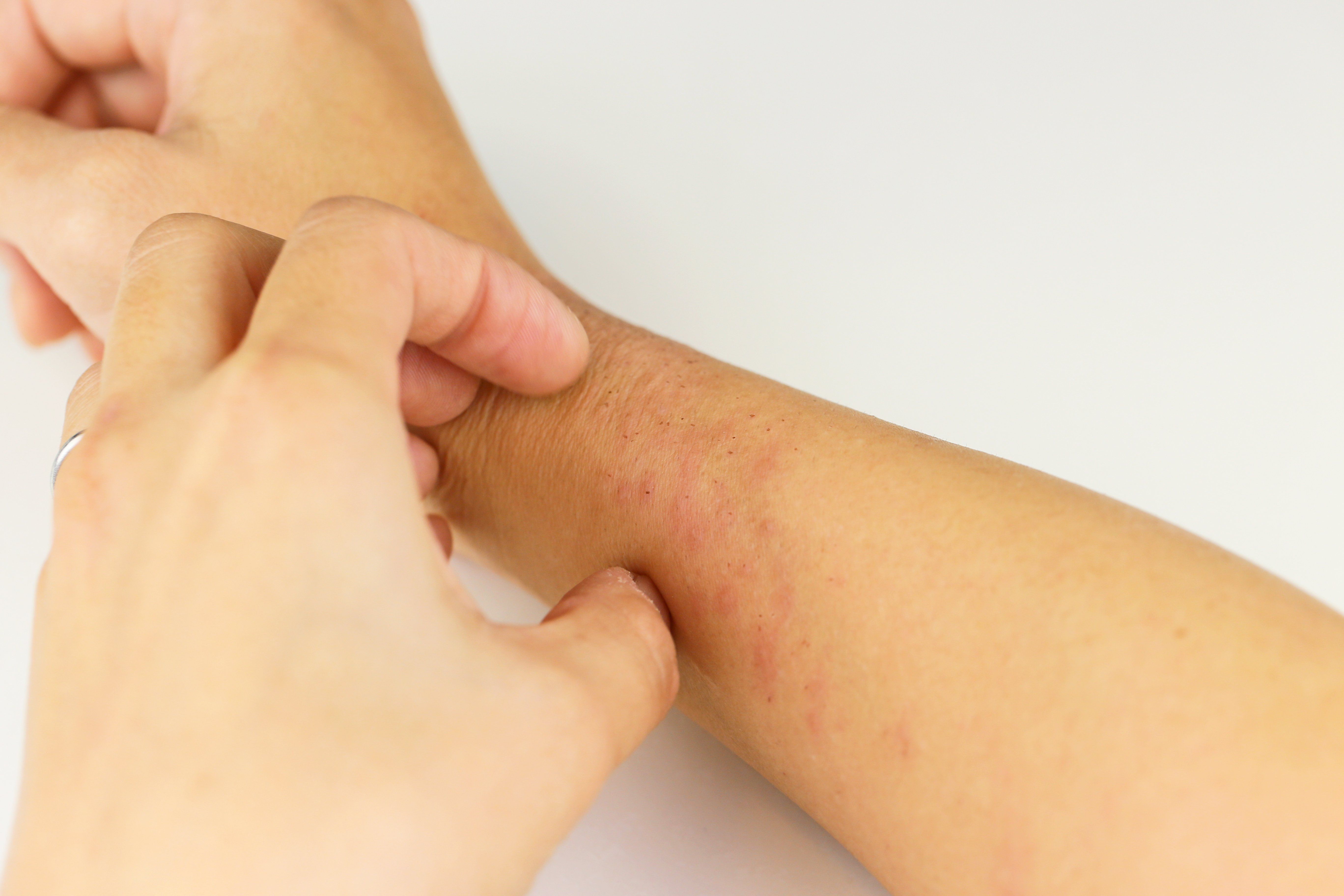
While neuropathic pain is a focus of research and drug development, the same cannot be said for neuropathic itch.
While neuropathic pain is a focus of research and drug development, the same cannot be said for neuropathic itch. (kelly marken - stock.adobe.com)

While neuropathic pain is a focus of research and drug development, the same cannot be said for neuropathic itch. As a result, the underlying mechanisms of neuropathic itch are poorly understood, diagnosis is challenging, and treatment options are limited.
Since inflammatory cutaneous signs like edema and erythema are characteristic of dermatological itch, and neurogenic inflammation can cause these signs in neuropathic itch as well, dermatologists should understand the underlying pathophysiology.
Neuropathic itch is the result of excess peripheral firing or dampened central inhibition of itch pathway neurons and a symptom of the same central and peripheral nervous system disorders that cause neuropathic pain, such as sensory polyneuropathy, radiculopathy, herpes zoster, stroke, or multiple sclerosis, according to Martin Steinhoff, M.D., M.Sc., department of dermatology and venereology, Hamad Medical Corporation, Doha, Qatar, who authored an article review that was published in The Lancet Neurology1.
The two conditions can occur simultaneously; however, unlike pain, itch is only felt in the skin or mucosa lining the body’s entrances.
Concomitant sensory loss and gain of function is observed in both neuropathic pain and itch, so a better understanding of the neuroanatomical and pathophysiological similarities and differences between the two conditions might identify existing but underused and novel treatment targets, he notes. To date, standardized case definitions to diagnose and differentiate the subforms of neuropathic itch and validated questionnaires to track symptoms are limited.
Diagnosis is complicated by the fact that different forms of neuropathic itch exist, such as focal vs. widespread and peripheral eral vs. central, as well as the fact that scratch-induced skin lesions can be mistaken as a primary (e.g., evidence of insect infestation) rather than a secondary symptom.
COMMON PRESENTATIONS
Small fibre peripheral neuropathy is one of the most common presentations of neuropathic itch and should be considered when patients present with unexplained chronic itch or scratch injuries in the length-dependent pattern on the limbs. It usually starts in the feet and progresses proximally in a lengthdependent pattern that sometimes also involves the hands.
Small fibre peripheral neuropathy occurs in approximately 40% of cases of fibromyalgia, and generalized axonopathies trigger more than half of all neuropathic itch presentations.
Focal mononeuropathies or oligoneuropathies that damage the small fibres within spinal nerves, plexi, or nerve roots are the major cause of focal neuropathic itch, Dr. Steinhoff writes. Itchy patches, which correspond to the cutaneous distribution of the damaged nerves or root, are most common on the head, upper torso, or arms, and are less common below the waist.
Compressive radiculopathy due to lateral spinal stenosis is the most common cause of brachioradial pruritus (the more distal patches), particularly after midlife. The second most common cause of truncal radicular neuropathic itch is herpes zoster, particularly at cervical and upper thoracic levels. Diabetic microvasculopathy should also be considered in patients with focal truncal neuropathic itch, as this requires prompt initiation of disease-specific treatment.
Ganglionopathies (neuronopathies) cause non-lengthdependent itch or itchy patches, and other sensory and radicular symptoms, neuropathic pain and proprioceptive ataxia. They can relate to cancer (particularly small-cell lung cancers), infection, or autoimmune disease, and imaging with contrast or cerebrospinal fluid analysis can help confirm these diagnoses.
Post-ganglionectomy ulcers were often misdiagnosed as being caused by deprivation of axonally transported nutritive trophic factors (trigeminal trophic syndrome), but then excessive and often painless scratching was recognized as the correct cause, Dr. Steinhoff writes. The para-midline nasal or the cheek is the characteristic location of the lesion after trigeminal ganglion or lower root injury, and the tip of the nose is usually spared. Non-trigeminal facial neuropathic itch can indicate lesions of the nervus intermedius of the cranial nerves VII or IX, or of the cervical spinal nerves C1 or C2, he adds. Herpes zoster is the most common cause of cranial neuropathic itch, and the forehead and anterior scalp are most commonly affected.
In diseases of the central nervous system, any type of lesion of the itch pathways in the spinal cord or brain can cause somatotopic neuropathic itch, including stroke, intramedullary neuromyelitis optica, intramedullary tumors, transverse myelitis, and spinal cord injury. Opioids and brain infections, such as Creutzfeldt-Jakob disease, can induce neuropathic itch, and neuropathic itch can present alone or with other symptoms such as colocalising sensory loss or weakness suggesting a neurologic origin.
The time between the onset of neuropathic itch or neuropathic pain can range between a few months to a few years after an event like a stroke, Prof. Steinhoff writes.
DIAGNOSTIC TOOLS
“Diagnosis is based primarily on recognizing clinical characteristics of specific neuropathic itch syndromes and appropriate confirmatory testing (e.g., for herpes zoster, diabetes),” Dr. Steinhoff says.
There are few diagnostic tools for neuropathic itch, and those emerging for chronic itch still require fine tuning for applicability to syndromes, he points out. Formal sensory testing (e.g., the Von Frey monofilament test) can highlight alloknesis and areas to treat topically, and measurement of epidermal nerve fibre density in PGP9·5-immunolabeled punch biopsies from the itchy skin, electrodiagnostic testing, and imaging can help to identify and localize causal neural injuries, Dr. Steinhoff writes. Autonomic testing, particularly the quantitative sudomotor axon reflex test, is useful for diagnosing small fibre peripheral neuropathy.
“Chronic itch questionnaires might improve diagnosis and enable the course of pruritus to be monitored,” he says. For example, the validated Massachusetts General Hospital Small-Fiber Symptom Survey captures “skin that itches for no reason.”
“If no dermatological or neurological causes for chronic itch can be identified, psychiatric conditions (e.g., trichotillomania, somatoform disorder) should be considered, particularly in patients with severe scratch injury,” he adds. “Rarely, a patient with no known physical cause for itch can become fixated on concerns over insect infestation (e.g., Ekbom syndrome or Morgellons disease, also termed delusional parasitosis).”
CURRENT MANAGEMENT APPROACHES
No specific therapies for neuropathic itch have been approved, and current treatment, such as opioids or antidepressants, is often based on clinical experience and trials. Antihistamines are prescribed for all forms of itch but are largely ineffective for neuropathic itch, Dr. Steinhoff writes; however, sedating antihistamines might be beneficial for improving sleep, reducing nocturnal scratching, and soothing scratch-induced skin inflammation.
“Treating scratch-induced lesions is important to reduce further itching, infection, scarring, and disfiguration,” he emphasized. Trimming fingernails or wearing gloves to bed can reduce scratching damage during sleep, and thermoplastic mesh coverage can improve wound healing by impeding further scratching.
“Cognitive behavioural therapy, physiotherapy, or meditation can also help people resist the urge to scratch,” Dr. Steinhoff writes. “Many patients benefit from occlusive therapy, where itchy patches are covered to reduce visual cues to scratch… Occlusive therapy physically protects the skin from further scratching and sun damage, and enhances penetration of topical therapies.”
Tightly bandaging itchy areas can provide relief by augmenting the inhibitory tone in the dorsal horn of the spinal cord, he notes, and if neuropathic itch is caused by compression of spinal nerves or roots, weight loss, physiotherapy, or other treatments to reduce nerve deformation may be of benefit.
In terms of pharmacological therapy, topical therapies should be considered first for peripheral neuropathic itch because they have few or no sideeffects. Topical local anesthetics are only effective for a few hours, and topical glucocorticosteroids are ineffective against primary causes of neuropathic itch but can soothe the associated inflammation that increases itch.
Daily subcutaneous injection of anesthetics near an injured superficial sensory nerve can provide effective short-term relief, and some patients can be taught to self-administer these.
Low-dose (0.025 – 0.1%) topical capsaicin has been used widely to treat neuropathic itch, Prof. Steinhoff highlights, but it requires reapplication four to five times a day. The first applications induce neurogenic inflammation and can cause pain, as well as worsen the itch. A few case reports suggest a higher concentration (8%) capsaicin patch benefits patients with brachioradial pruritus, notalgia paraesthetica, HIV-associated peripheral neuropathic pain, or diabetes-associated peripheral neuropathic pain, and subcutaneous injections of botulinum toxin A have shown efficacy in neuropathic itch, he notes.
Oral medications should be considered for patients with widespread neuropathic itch or refractory to topical treatments.
Systemic sodium channel blockers, such as carbamazepine and oxcarbazepine are the first-line choice, as they calm the ectopic neuronal firing. Gabapentin and pregabalin are commonly prescribed for patients with brachioradial pruritus and appear effective at similar concentrations as used for neuropathic pain.
Reference
Steinhoff M, Schmelz M, Szabó IL, Oaklander AL. Clinical presentation, management, and pathophysiology of neuropathic itch. Lancet Neurol. 2018;17(8):709-720. https://www.thelancet.com/journals/laneur/article/PIIS1474-4422(18)30217-5/fulltext.


