- Case-Based Roundtable
- General Dermatology
- Eczema
- Chronic Hand Eczema
- Alopecia
- Aesthetics
- Vitiligo
- COVID-19
- Actinic Keratosis
- Precision Medicine and Biologics
- Rare Disease
- Wound Care
- Rosacea
- Psoriasis
- Psoriatic Arthritis
- Atopic Dermatitis
- Melasma
- NP and PA
- Skin Cancer
- Hidradenitis Suppurativa
- Drug Watch
- Pigmentary Disorders
- Acne
- Pediatric Dermatology
- Practice Management
- Prurigo Nodularis
- Buy-and-Bill
Publication
Article
Dermatology Times
Leg ulcers that require punch biopsies
Author(s):
Victor Desmond Mandel, M.D. and colleagues describe a reconstruction strategy for chronic infected neoplastic ulcers of the lower extremities using a dermal matrix and a unique postoperative dressing protocol.
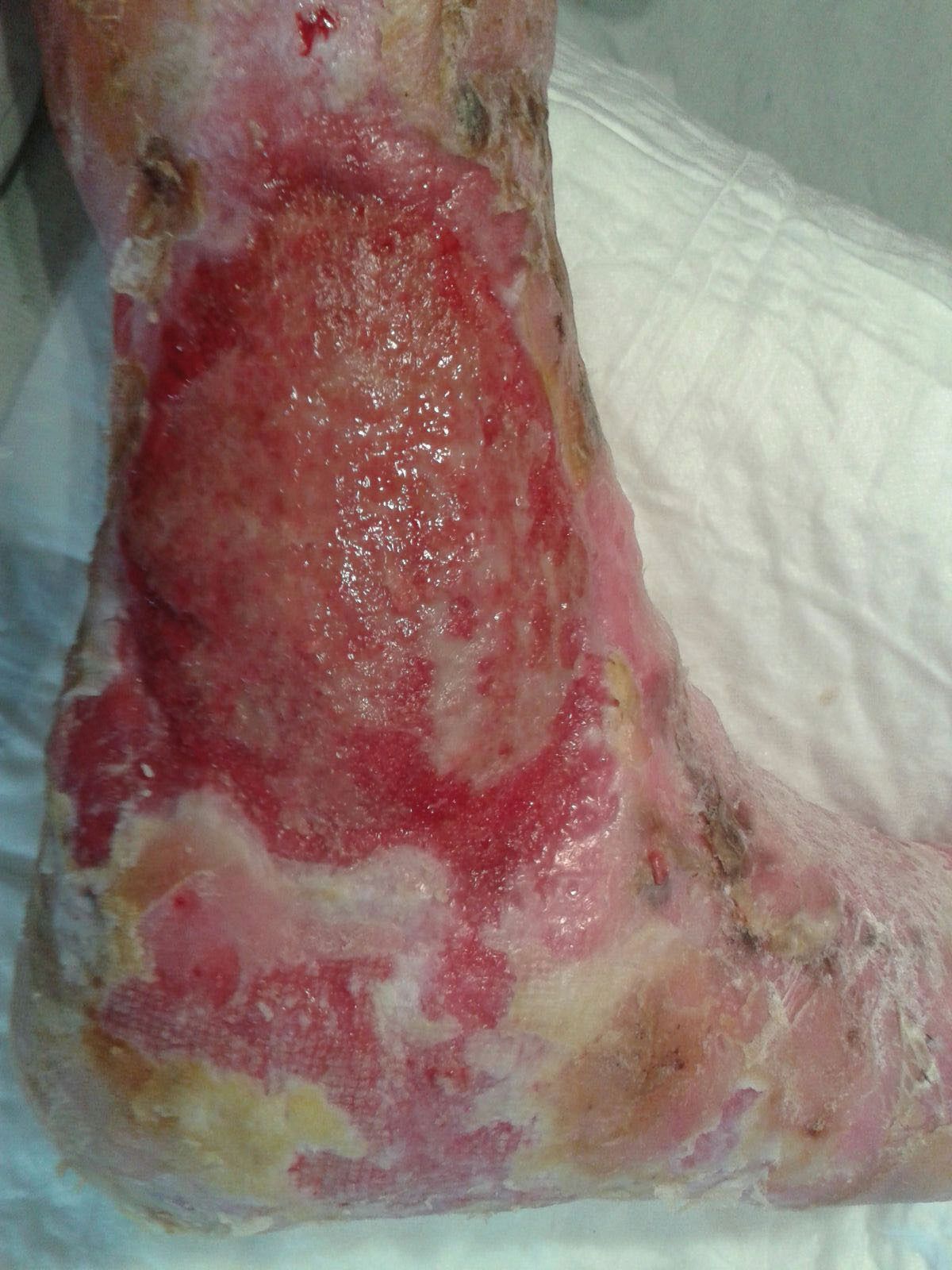
Non-healing exophytic lesion on the right malleolus suggestive for squamous cell carcinoma. (Photos courtesy of Dr. Victor Desmond Mandel)
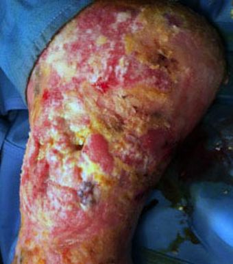
Image A. Photo courtesy of Dr. Victor Desmond Mandel.
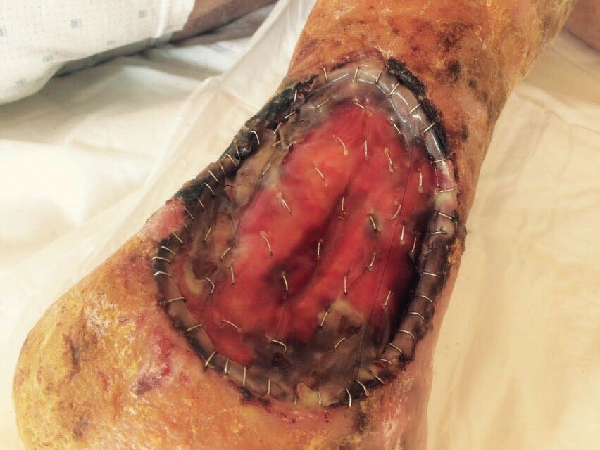
Image B. Photo courtesy of Dr. Victor Desmond Mandel.
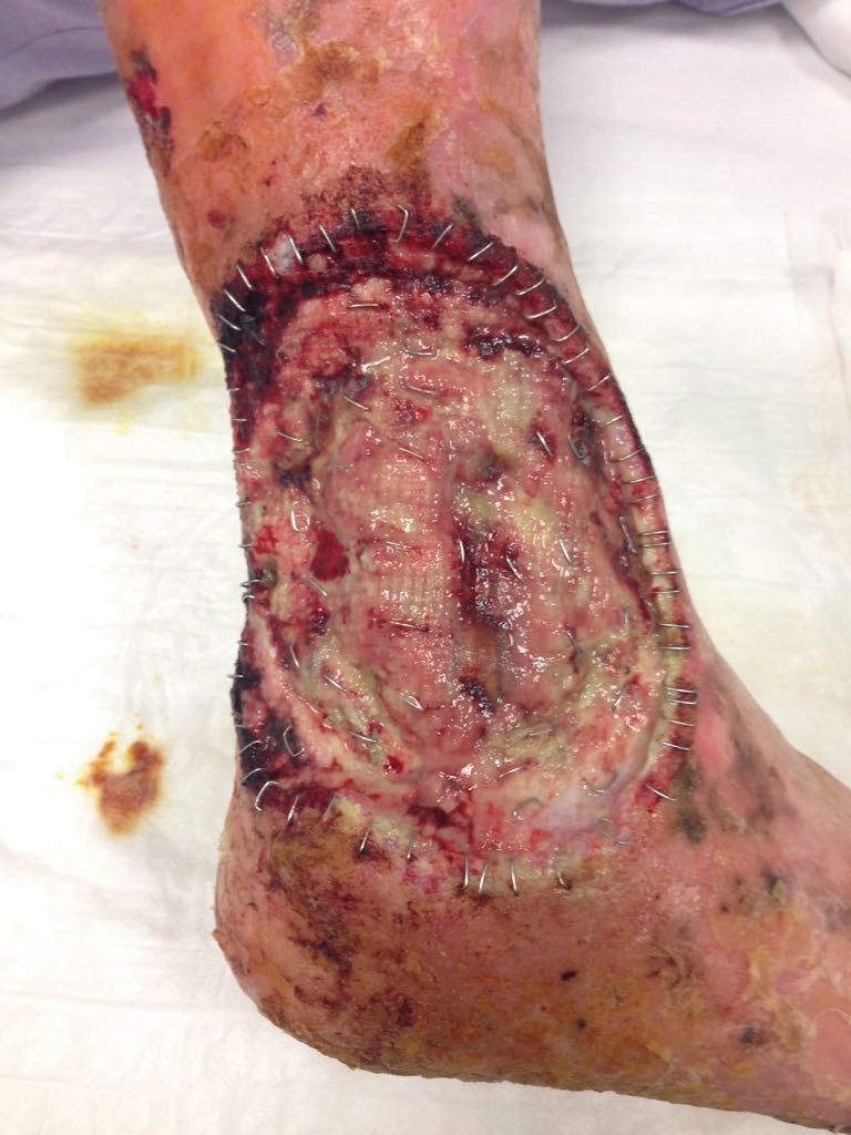
Image C. Photo courtesy of Dr. Victor Desmond Mandel.
Leg ulcers can be caused by vasculitis, pressure sores, inflammatory diseases, traumatic injuries and cutaneous neoplasmsâwhich are often misdiagnosed as skin ulcers, write Lidia Sacchelli and colleagues in the September 19 online issue of the JournalDermatopathology (DOI:10.1159/000491922).
“We believe that clinicians should be aware of the importance of an early and correct diagnosis of cutaneous neoplasms underlying chronic leg ulcers. This could avoid diagnostic delay and guarantee the best therapeutic approach to each patient,” they wrote.
Lesions that grow, spread or are pigmented may require a biopsy and histologic study. If the wound doesn’t heal within three months of treatment, that may indicate the presence of a neoplasm requiring a skin biopsy and histologic study.
In the Sept. 11 issue of the Journal of Wound Care (doi:10.12968/jowc.2018.27.9.558), Victor Desmond Mandel, M.D., and colleagues from the oncology and regenerative medicine unit of the University of Modena and Reggio Emilia, Italy, describe a reconstruction strategy for chronic infected neoplastic ulcers of the lower extremities using a dermal matrix.
In one of two cases featured in the case report, they describe a 46-year-old male patient with laminin-332-deficient non-Herlitz junctional epidermolysis bullosa (JEB-nH).
Four months prior, the patient was admitted to the hospital with a 10x8 cm non-healing exophytic lesion on the right malleolus with visible scleroatrophic areas, erosions and hyperkeratosis, but no signs of acute infection.
A biopsy confirmed squamous cell carcinoma. A surgical excision was conducted on the exophytic lesion followed by reconstruction with a dermal matrix and silicone.
After three days, the matrix became infected (image A) and was removed. The surgeons then performed a single surgical hydrodebridement of the wound bed and a new graft with a dermal matrix (image B), followed by a post-operative treatment regime consisted of several layers of dressings in order to prevent surgical site infection. In this case a skin graft was impossible to perform because of the genetic condition. Image C shows the results achieved one month after reconstruction with dermal matrix and the post operative dressing.
“In our patients, the use of dermal matrix in conjunction with our dressing protocol resulted in complete wound healing. We achieved perfect engraftment of the matrix, almost complete restoration of the missing volume and good quality of grafted skin,” Dr. Mandel and colleagues wrote.
