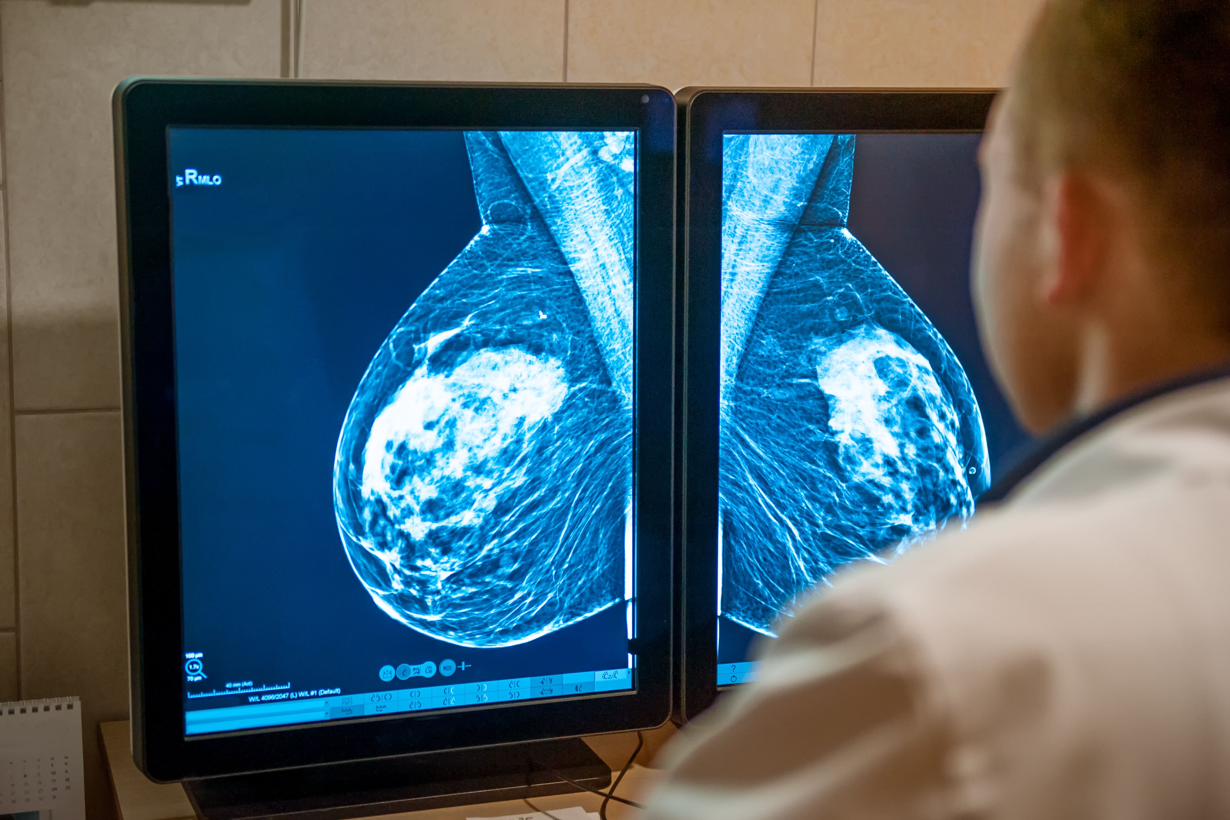
October is Breast Cancer Awareness Month, and although it is critical for oncologists to have a thorough understanding of the condition’s impact on the entire body, dermatology clinicians play an important role in a multidisciplinary care plan. Skin conditions can be associated with the presentation, progression, and treatment of breast cancer. “Dermatologists may be the first to identify a breast cancer diagnosis, as a subset of patients first present with direct extension of an underlying tumor or with a cutaneous metastasis,” according to Milam et al.1
Key Takeaways
- Dermatologists can be the first to identify a breast cancer diagnosis and play an integral part of a multidisciplinary care plan for patients.
- A variety of skin conditions can arise from the surgical and radiation treatments of breast cancer.
- Beth McLellan, MD, presented 4 cases of adverse effects of breast cancer treatments earlier this year at AAD.
Surgical treatment can bring a variety of skin conditions to the table including postoperative lymphedema, soft tissue infections, seromas, pyoderma gangrenosum, and scarring disorders. Radiation treatment on the breasts can often result in skin changes from mild dermatoses to disfiguring ulcers, fibrosis, and necrosis. More rare conditions can include angiosarcoma, radiation-induced morphea, lichen planus, and postirradiation pseudosclerodermatous panniculitis.
Adverse Effects of Treatments
Beth McLellan, MD, dermatology professor at the Albert Einstein College of Medicine in Bronx, New York, has a special interest in supportive oncodermatology. She presented during the session “Behind the Bra: What Dermatologists Should Know About Diseases of the Breast” earlier this year at the American Academy of Dermatology Annual Meeting in New Orleans, Louisiana. Her focus was on adverse effects of breast cancer treatments. She presented 4 cases and several pearls for her colleagues in dermatology.2
Case 1: Woman aged 31 years with invasive intraductal carcinoma
This patient’s cancer treatment included neoadjuvant chemotherapy and then bilateral mastectomy with reconstruction. She began adjuvant radiation therapy with 50.4 Gy to chest wall and lymph nodes. Radiation has been an effective tool for treating cancer for more than 100 years, and nearly 90% of patients will develop radiation dermatitis.3
McLellan’s radiation dermatitis pearls include considering mometasone and bacterial decolonization for prophylaxis. She also suggests always checking for secondary infection and treating as needed.
Case 2: Woman aged 61 years with history of invasive ductal carcinoma and psoriasis
This patient had a skin-sparing right mastectomy with implant reconstruction and axillary dissection along with chemotherapy, radiation therapy, and anastrozole. She visited her dermatologist with a 7-day history of itchy rash in the radiation field extending to the neck. Her diagnosis was eosinophilic, polymorphic, and pruritic eruption associated with radiotherapy, which is most common after cervical and breast cancer. It can mimic bullous pemphigoid and dermatitis herpetiformis. It often presents outside of radiated areas, and the risk appears to be dose dependent (mean, 30 Gy).
McLellan shared that this patient’s rash was cleared with triamcinolone 0.1% ointment without recurrence. Pearls to treat eosinophilic, polymorphic, and pruritic eruption associated with radiotherapy include considering the diagnosis in a patient with a widespread eruption and history of radiation and treating with phototherapy, topical steroids, or systemic steroids.
Case 3: Woman aged 62 years with stage IIA, lymph node–negative infiltrating ductal carcinoma
She was treated with a left breast partial mastectomy, radiation therapy, and chemotherapy (AC-T; doxorubicin hydrochloride [Adriamycin], cyclophosphamide, and paclitaxel [Taxol]). Three weeks after radiation, the patient experienced redness and swelling of the left breast and underwent 3 courses of cephalexin (Keflex) without improvement. Multiple skin biopsies showed radiation changes and fibrosis. The diagnosis was postirradiation morphea, and she saw improvement with pentoxifylline and vitamin E.
McLellan explained that postirradiation morphea often is mistaken for mastitis and that there is a 20% to 30% spread beyond the radiation field. Treatment options include topical and intralesional corticosteroids, methotrexate, pentoxifylline with vitamin E, phototherapy (UV-A1 or narrowband UV-B), surgical excision, and/or physical therapy. Postirradiation morphea may need several biopsies.
Case 4: Woman aged 45 years with history of left breast cancer
She underwent neoadjuvant chemotherapy and then a skin-sparing bilateral mastectomy with tissue expander placement, adjuvant radiation, and tamoxifen. Six months after surgery, she was referred for an asymptomatic rash on the right lateral breast. Previously, the patient has not been responsive to emollients or trimethoprim-sulfamethoxazole. She was given fluocinonide 0.05% ointment. The rash improved but recurred, requiring a shave biopsy. The shave biopsy led to a diagnosis of nummular dermatitis of the reconstructed breast.
Less than 3% of patients develop this condition with variable timing. In addition, 42% of cases have a periwound and 67% present nonperiwound.4 Nummular dermatitis of the reconstructed breast pearls to keep in consideration include the following: It is not always itchy or occurring after reconstruction; biopsies are essential to rule out cutaneous metastases; scrape to rule out fungus; topical steroids usually trigger a response.
References
1. Milam EC, Rangel LK, Pomeranz MK. Dermatologic sequelae of breast cancer: from disease, surgery, and radiation. Int J Dermatol. 2021;60(4):394-406. doi:10.1111/ijd.15303
2. Mansh M, Markova A, Murase J, et al. Behind the bra: what dermatologists should know about diseases. Presented at: American Academy of Dermatology 2023 Annual Meeting; March 17-21, 2023; New Orleans, LA.
3. Siegel R, Ma J, Zou Z, Jemal A. Cancer statistics, 2014. CA Cancer J Clin. 2014;64(1):9-29.2014;64(5):364. doi:10.3322/caac.21208
4. Iwahira Y, Nagasao T, Shimizu Y, Kuwata K, Tanaka Y. Nummular eczema of breast: a potential dermatologic complication after mastectomy and subsequent breast reconstruction. Plast Surg Int. 2015;2015:209458. doi:10.1155/2015/209458


