- Case-Based Roundtable
- General Dermatology
- Eczema
- Chronic Hand Eczema
- Alopecia
- Aesthetics
- Vitiligo
- COVID-19
- Actinic Keratosis
- Precision Medicine and Biologics
- Rare Disease
- Wound Care
- Rosacea
- Psoriasis
- Psoriatic Arthritis
- Atopic Dermatitis
- Melasma
- NP and PA
- Skin Cancer
- Hidradenitis Suppurativa
- Drug Watch
- Pigmentary Disorders
- Acne
- Pediatric Dermatology
- Practice Management
- Prurigo Nodularis
- Buy-and-Bill
Publication
Article
Dermatology Times
3D imaging technique measures severity of atrophic acne scars
Author(s):
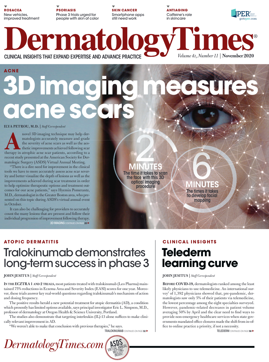
There is a dire need to improve the clinical tools dermatologists use to assess the severity of acne scars. This 3D imaging technique may help, according to a recent study presented at the 2020 virtual American Society for Dermatologic Surgery (ASDS) meeting.
A novel 3D imaging technique may help dermatologists accurately measure and grade the severity of acne scars as well as the aesthetic improvements achieved following scar therapy in atrophic acne scar patients, according to a recent study presented at the American Society for Dermatologic Surgery (ASDS) Virtual Annual Meeting.
Clinicians currently have several validated measuring and grading tools at their disposal to help them evaluate the severity of acne scarring, including the Global Aesthetic Improvement Scale, ECCA, and the Goodman and Baron grading scales. But these techniques aren’t perfect; they lack objectivity, do not reflect 3D morphology of scars, are easily affected by ambient light and can be time-consuming. begging the need for more accurate and precise scar evaluation and grading tools.
Hyemin Pomerantz, MD

“There is a dire need for improvement in the clinical tools we have to more accurately assess acne scar severity, better visualize the depth of lesions as well as the improvements achieved during scar treatment in order to help optimize therapeutic options and treatment outcomes for our acne patients,” says Hyemin Pomerantz, M.D., dermatologist in the Greater Boston area, who presented on this topic during ASDS.
It can also be challenging for providers to accurately count the many lesions that are present and follow their individual progression of improvement following therapy. Atrophic acne scars are notoriously challenging to treat and as such, aesthetic improvements following scar therapy are typically scrutinized for visible, measurable improvements before patients are willing to undergo the necessary repeated treatments.
“Acne scar therapy is usually not a one-off treatment and the vast majority of patients will typically undergo multiple procedures to achieve cosmetic improvement of their acne scars,” Dr. Pomerantz adds.
Dr. Pomerantz and Roy G. Geronemus, M.D., director of the Laser & Skin Surgery Center of New York, investigated the reliability of a novel 3D high-resolution, stereoscopic optical imaging system (Cherry Imaging, Cherry Imaging, Ltd), as an alternative clinical tool for measuring atrophic scars severity on the face. The prospective study included six male and five female patients aged 18 years or older (average age 34 years) with Fitzpatrick skin types ranging from I-VI and who had atrophic facial scars for an average of 11 years.
Three operators scanned the subjects’ faces at two different times. Image software calculated the volume of atrophic acne scars and index volume (volume normalized by surface area of acne scars) on the forehead and left and right cheeks. ECCA grading was recorded by a dermatologist. A two-way ANOVA test was used to determine intraclass correlation coefficient (ICC) among the three operators to examine inter-rater reliability and ICC at two different times for each operator to examine intra-rater reliability. The volume from the 3D imaging and ECCA scores were also correlated.
Results showed that the volume and index volume of atrophic acne scars on the forehead, left cheek, and right cheek, separately, were consistent independent of operators (ICC range 0.72 to 0.92). The ICC of volume and index volume of the scars on the areas of the forehead, left and right cheeks combined was 0.88 and 0.89, respectively. Intra-rater reliability was consistent with average ICC’s of the three operators showing 0.95 for atrophic acne scar volume and 0.90 for index volume. Data also showed that Pearson correlation with ECCA scores were 0.22 and 0.48 for volume and index volume, respectively.
One of the main advantages of this grading system is that it does not rely on the clinical experience of the evaluator, Dr. Pomerantz notes.
“The procedure could also be easily relegated to office staff as it is non-invasive and standardized to eliminate any subjectivity in evaluation,” says Dr. Pomerantz. “Moreover, it also precisely gives you the total volume of acne scars in the face, which is something more tangible that patients can better understand effects of their treatment course.”
Similar to a GPS system, the innovative device uses a stereoscopic technology to lock-on and measure precise 3D coordinates of the targeted area. Akin to facial mapping, the system creates an image of the face upon which the exact placement and measurement parameters of the acne scars are superimposed. According to Dr. Pomerantz, it takes approximately 2 minutes to scan the entire face and another 6 minutes to develop the facial mapping image, sparing much valuable time for the operator and patients alike.
There is a gap in measuring the progress of a number of dermatologic conditions during a patient’s therapeutic course, and lesions in 3D configuration like scars and skin laxity are challenging to measure and quantify, notes Dr. Pomerantz.
“This system offers clinicians and their patients an opportunity to accurately and objectively discuss standardized images and results from treatment, instead of viewing before and after treatment images that can be interpreted differently because of subjectivity,” she says. “Having an easy-to-use 3D modality at our disposal to quickly quantify the therapeutic outcomes in our patients is the future of state-of-the-art acne scar therapy.”
Disclosures: Dr. Pomerantz is a consultant for Accure and Avava. The study was supported by ASDS Frederic S. Brandt, MD, Innovations in Aesthetics Fellowship grant.
References:
Pomerantz, H. “Validation of Reliability of 3D Imaging for Objective Measurement of Facial Atrophic Acne Scars.” American Society for Dermatologic Surgery Virtual Annual Meeting. October 11, 2020.


