- Case-Based Roundtable
- General Dermatology
- Eczema
- Chronic Hand Eczema
- Alopecia
- Aesthetics
- Vitiligo
- COVID-19
- Actinic Keratosis
- Precision Medicine and Biologics
- Rare Disease
- Wound Care
- Rosacea
- Psoriasis
- Psoriatic Arthritis
- Atopic Dermatitis
- Melasma
- NP and PA
- Skin Cancer
- Hidradenitis Suppurativa
- Drug Watch
- Pigmentary Disorders
- Acne
- Pediatric Dermatology
- Practice Management
- Prurigo Nodularis
- Buy-and-Bill
News
Article
Vitiligo Case Report: A 9-Year Old Patient With White Macules, Halo Nevus
Author(s):
Jordan Bui and Bernard A. Cohen, MD, review a case of a young patient with vitiligo.
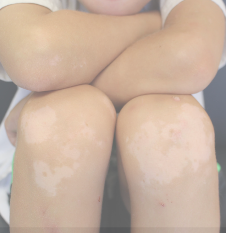
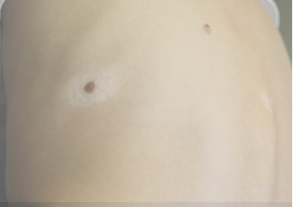
A 9-year-old Asian boy presents for evaluation of white macules on his hands, elbows, knees, and legs (Figures 1,2). There is also an oval symmetric halo of depigmentation around a mole on his back. The patient is very active and spends a lot of his time outdoors playing soccer. The patient’s parents first noted white macules on his trunk and extremities, and his lesions have spread rapidly over 2 months particularly in areas of trauma where his soccer padding overlie the knees. The lesions were most noticeable in the summer when tanning increased the contrast between the involved and uninvolved areas of his body. The lesions have no associated pruritis or pain.
Figure 1: Vitiligo on extensor surfaces

Aside from expressed anxiety by the patient, review of other systems and laboratory data are unremarkable. There is no other significant medical history in the patient or family. Physical exam with dermoscopy demonstrated telangiectasias and perifollicular pigmentation and Wood’s lamp demonstrated sharply demarcated areas of depigmentation. This patient’s hypopigmented lesions and halo nevus are consistent with non-segmental generalized vitiligo.
Vitiligo is a chronic disorder of pigmentation with a prevalence of 0.76% in the U.S. and 25% of cases occurring before the age of 10 (Farajzadeh). The pathology of vitiligo is not entirely known but suspected to be due to a combination of autoimmune, genetic, and environmental factors. Vitiligo is highly associated with autoimmune diseases such as Hashimoto’s thyroiditis, type 1 diabetes mellitus, alopecia areata, rheumatoid arthritis, and inflammatory bowel disease. Several cytokines and cytotoxic T lymphocytes have been implicated in the destruction of melanocytes, causing vitiligo. Additionally, there is a suggested genetic predisposition with vitiligo, with 20% of affected individuals having relatives with vitiligo as well(Alkhateem). Environmental factors such as physical stress, recurrent infections, and malnutrition may be triggers for disease onset. As in our case, sun exposure is another environmental trigger for vitiligo. Vitiligo development in areas of repeated trauma due to friction, scratching, pressure, or other irritants is known as the Koebner phenomenon. The patient’s presenting halo nevus has a 26% incidence in children with non-segmental vitiligo and more often presents in children with onset of vitiligo younger than 12 years of age (Cohen). In adolescents, development of halo nevi is not unusual and is a benign finding that does not alter the severity of disease.However in older adults, development of halo nevi may be rarer and can warrant skin checks for other malignancies.
The patient was started on oral prednisolone 5mg daily, topical calcipotriol, and 0.1% topical mometasone furoate to facilitate stabilization of vitiligo. Combination therapy is preferred and more successful than monotherapy in repigmentation and cessation of hypopigmentation spread. After three months of steroid treatment, repigmentation was slow and did not seem responsive. The patient then underwent targeted UVB phototherapy and trial of topical calcineurin inhibitor (tacrolimus) with greater success and repigmentation within six months.
Figure 2: Halo nevus - oval area of hypopigmentation surrounding nevus on back.

In younger children it is important to consider how vitiligo can affect quality of life. Children with vitiligo report higher rates of depression, greater self-consciousness, more trouble making friends, and more frequent bullying. This experience is most notably associated with decreased quality of life in children ages 6-11, when they develop a sense of identity and community during school (Silverberg). Thus, psychological referral should also be considered as part of treatment in conjunction with repigmentation therapies. Because of the chronicity of vitiligo, patients may require intermittent maintenance treatment with topical steroids, topical tacrolimus, or phototherapy for long term management. There is no associated increased risk of melanoma or other skin cancers in patients with vitiligo.
It is important to be mindful of other hypopigmented and depigmented diseases that may present similarly to vitiligo. Nevus depigmentosus presents at birth or the first years of life as a circumscribed hypopigmented or depigmented solitary lesion that shows little change over time. Pityriasis alba presents as multiple asymptomatic hypopigmented, mildly scaling patches on sun-exposed area. Tinea versicolor is a superficial yeast infection that presents as hypopigmented, pale macules on sun-exposed areas such as the upper trunk and chest with a dry scale. The scaling and textural changes present in some of these conditions are not associated with vitiligo.
References
- Farajzadeh S, Khalili M, Mirmohammadkhani M, Paknazar F, Rastegarnasab F, Abtahi-Naeini B. Global clinicoepidemiological pattern of childhood vitiligo: a systematic review and meta-analysis. BMJ Paediatr Open. 2023;7(1):e001839. doi:10.1136/bmjpo-2022-001839
- Alkhateeb A, Fain PR, Thody A, Bennett DC, Spritz RA. Epidemiology of vitiligo and associated autoimmune diseases in Caucasian probands and their families. Pigment Cell Res. 2003;16(3):208-214. doi:10.1034/j.1600-0749.2003.00032.x
- Cohen BE, Mu EW, Orlow SJ. Comparison of Childhood Vitiligo Presenting with or without Associated Halo Nevi. Pediatr Dermatol. 2016;33(1):44-48. doi:10.1111/pde.12717
- Silverberg JI, Silverberg NB. Quality of life impairment in children and adolescents with vitiligo. Pediatr Dermatol. 2014;31(3):309-318. doi:10.1111/pde.12226


