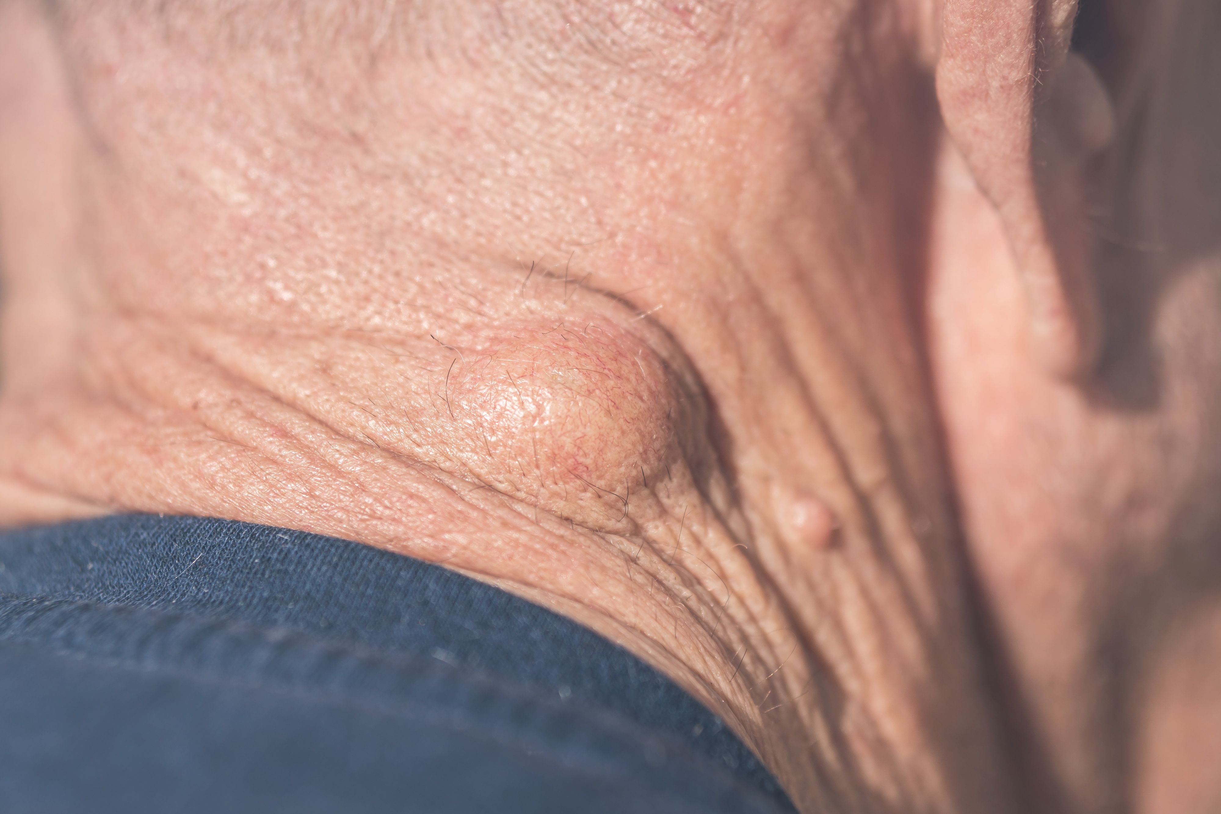
Lesion types included:
- Epidermoid cysts (n=225)
- Lipomas (n=77)
- Pilomatrixomas (n=25)
- Hemangiomas (n=21)
- Dermatofibromas (n=19)
- DFSPs (n=11)
- Neurofibromas (n=7)
- Leiomyomas (n=6)
News
Article
Author(s):
Indeterminacy of invisible subcutaneous lesions such as epidermoid cysts, lipomas, pilomatrixomas, hemangiomas, and DFSPs, can be decreased with HFUS use, according to investigators.
The use of high-frequency ultrasound (HFUS), when combined with traditional clinical examination, can improve diagnostic accuracy and determinacy of subcutaneous lesions, according to investigators behind a recent study1 published in Skin Research and Technology. Invisible lesions such as epidermoid cysts, lipomas, pilomatrixomas, hemangiomas, and dermatofibrosarcoma protuberans (DFSPs), may be more easily recognizable and diagnosable as a result of HFUS use.

According to investigators Miao et al, nonspecific presentation of invisible subcutaneous lesions may lead to delayed treatment, unnecessary biopsy, or the implementation of an incorrect treatment action plan. They sought to explore the use of ultrasound in identification and diagnosis of invisible lesions due to its balance of resolution and penetration, unlike other imaging modalities such as computed tomography and magnetic resonance imaging.
Investigators conducted the study, which was retrospective in nature, between December 2016 and July 2021. Patients who had visited the study’s dermatology department and had presented with an invisible subcutaneous lesion were eligible for participation. Additionally, they were required to have undergone HFUS evaluation of such lesions and have pathological results available for review following biopsy or surgery.
Patients with a prior treatment history for their subcutaneous lesions, who had hernias/deeply-muscular lesions/lesions of organic origin, or for whom pathological results were unclear, were excluded from participation.
In total, 355 patients between the ages of 5 and 86 years of age were enrolled in the study, amassing a total of 391 lesions.
Diagnosis of each lesion was determined by one clinician and further evaluated by a second clinician, who was blinded. HFUS imaging was conducted for each patient, and a final diagnosis was made through combining clinical examination findings and HFUS imaging findings.
As a result, the average accuracy of clinical diagnoses was 47.3%. Furthermore, 36.6% of lesions were indeterminantly diagnosed through clinical examination alone, while 16.1% were incorrectly diagnosed. Diagnostic accuracy was even lower in rarer subcutaneous lesions, totaling 13.5% accuracy in lesions such aspilomatrixoma, hemangioma, DFSP, dermatofibroma, neurofibroma, and leiomyoma.
When combining HFUS imaging with clinical examination, however, accuracy of diagnosis for non-rare subcutaneous lesions improved significantly in epidermoid cysts, lipomas, pilomatrixomas, hemangiomas, and DFSPs. In epidermoid cysts, accuracy had improved from 59.6% to 86.7% with the addition of HFUS. In lipomas, accuracy improved from 50.6% to 94.8% as a result.
There was, however, no significant difference in identification accuracy among dermatofibroma, neurofibroma, or leiomyoma.
“The combined diagnosis of HFUS and clinical examination can generally improve the diagnosis accuracy and decrease the number of unconfirmed cases of invisible subcutaneous lesions,” according to study authors Miao et al. “In detail, combined diagnosis can correctly diagnose more epidermoid cysts, lipomas, pilomatrixomas, hemangiomas, and DFSPs compared to clinical diagnoses. However, for some relatively rare cases in our study, such as dermatofibromas, neurofibromas, and leiomyomas, HFUS failed toprovide useful information.”
Reference