- General Dermatology
- Eczema
- Chronic Hand Eczema
- Alopecia
- Aesthetics
- Vitiligo
- COVID-19
- Actinic Keratosis
- Precision Medicine and Biologics
- Rare Disease
- Wound Care
- Rosacea
- Psoriasis
- Psoriatic Arthritis
- Atopic Dermatitis
- Melasma
- NP and PA
- Skin Cancer
- Hidradenitis Suppurativa
- Drug Watch
- Pigmentary Disorders
- Acne
- Pediatric Dermatology
- Practice Management
- Prurigo Nodularis
Article
Sweet's syndrome: Diagnosis not always straightforward
Author(s):
Sweet's syndrome is not always a straightforward diagnosis, especially when extracutaneous manifestations dominate the initial clinical picture. According to one expert, a careful history and an open mind are key in reaching a quick and accurate diagnosis.

Key Points

Extracutaneous manifestations of Sweet's syndrome are rare, though, and according to one expert, these atypical manifestations of the disease may at first perplex clinicians and may lead to confusion and diagnostic limbo.
"Rarely, you can have systemic involvement with Sweet's syndrome, such as joint effusions and pulmonary systems, without the initial presentation of the classic papular and nodular skin manifestations," says Eva R. Parker, M.D., at Southwest Dermatology, Orland Park, Ill.
Recently, Dr. Parker and colleagues admitted a 60-year-old female patient who presented with a diffuse rash, general malaise, fever and arthralgias, as well as a non-productive cough.
The patient reported that 10 days prior to presentation, she had an upper respiratory illness for which the treating physician at another center prescribed amoxicillin. After experiencing an initial improvement, the patient quickly deteriorated and got worse.
After presentation to Dr. Parker, the patient received a chest X-ray and a CAT scan, which showed a dense consolidation in her lower left lobe, confirming positive pulmonary findings.
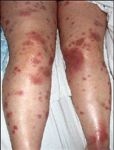
The patient slowly developed extensive, large erythematous-violaceous plaques extending on her extremities and trunk, as well as joint pain with effusions.
"Only a few of the lesions on the patient's arm were more typical of what you would see in Sweet's syndrome, with a pseudo-vesicular appearance.
"Therefore, we had a wide differential ranging from Sweet's syndrome, an infectious process, or a drug-related eruption such as erythema multiforme major," Dr. Parker says.
Biopsies
The first biopsy was inconclusive, showing a cellular infiltrate that was more granulomatous than neutrophilic.
The patient's condition deteriorated, leading Dr. Parker to perform a second biopsy the following day, as well as tissue cultures, a bronchoscopy, a bronchial-alveolar lavage and a lung biopsy.
The second set of skin lesion biopsies demonstrated a histologic picture much more typical of Sweet's syndrome with dense neutrophilic infiltrates.
The bronchial-alveolar lavage also showed an increased white blood count with a predominance of neutrophils.
Treatment course
"In Sweet's syndrome, the treatment of choice is steroids; however, this treatment could prove harmful if a systemic infection were present," Dr. Parker says.
Dr. Parker started the patient on high-dosage oral steroids, to which the patient responded dramatically. Within 12 hours of the treatment course, the patient's skin lesions began to clearly resolve, and by 48 hours, the lesions almost completely resolved, with only some remnant patchy erythema to be seen.
Etiology
Dr. Parker says it is good practice for clinicians to maintain a high level of suspicion for Sweet's syndrome when searching for an accurate diagnosis, especially in patients with fever, cutaneous neutrophilic infiltrates and pulmonary symptoms when no infectious etiology can be identified.
Disclosure: Dr. Parker reports no relevant financial conflicts.


