- Case-Based Roundtable
- General Dermatology
- Eczema
- Chronic Hand Eczema
- Alopecia
- Aesthetics
- Vitiligo
- COVID-19
- Actinic Keratosis
- Precision Medicine and Biologics
- Rare Disease
- Wound Care
- Rosacea
- Psoriasis
- Psoriatic Arthritis
- Atopic Dermatitis
- Melasma
- NP and PA
- Skin Cancer
- Hidradenitis Suppurativa
- Drug Watch
- Pigmentary Disorders
- Acne
- Pediatric Dermatology
- Practice Management
- Prurigo Nodularis
- Buy-and-Bill
Publication
Article
Dermatology Times
Rosacea commonly underdiagnosed
Author(s):
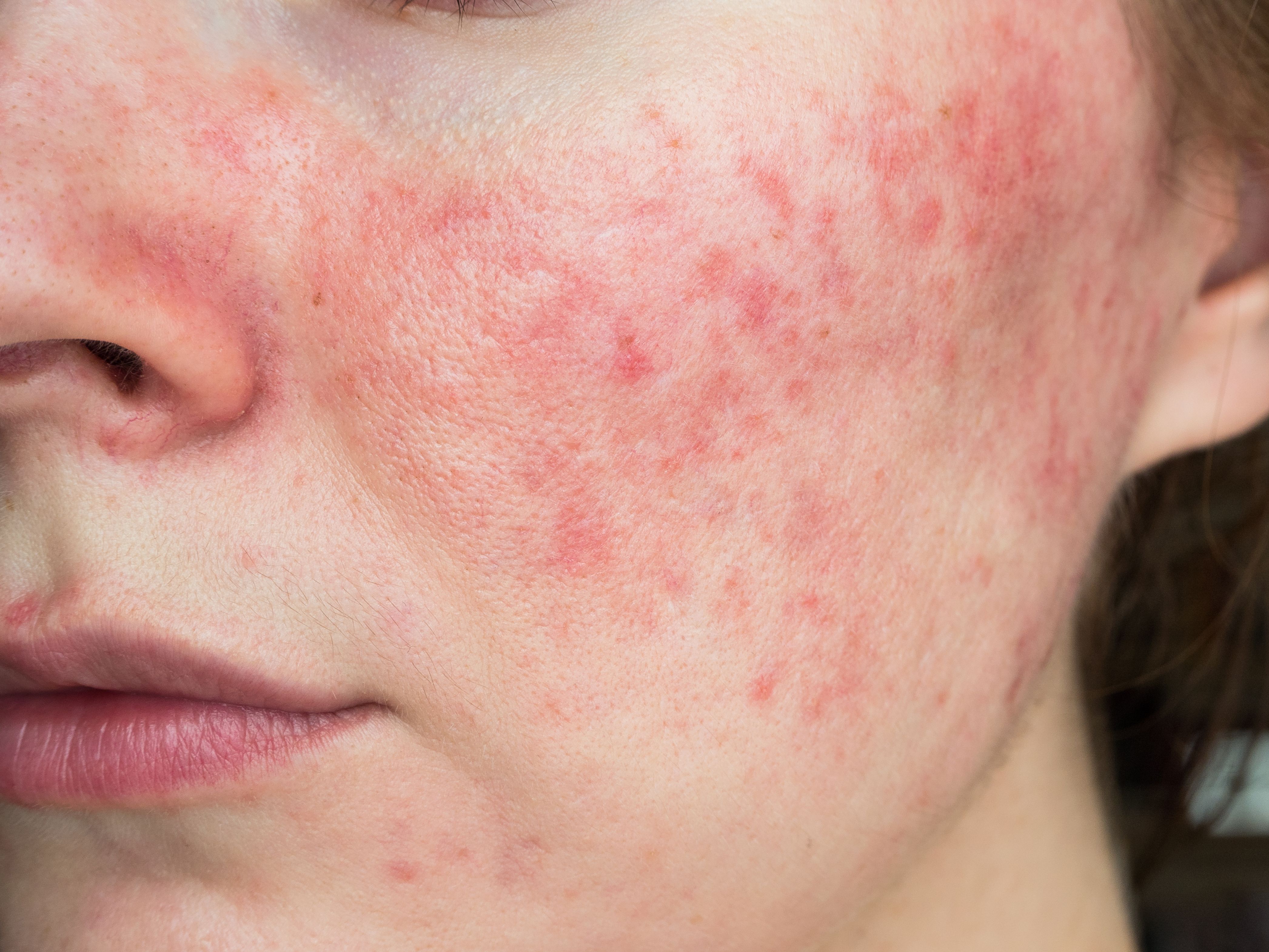
It's thought that rosacea affects about 10% of the population, but this estimation may not be completely accurate as the condition is often underdiagnosed – especially in individuals with darker skin types.
Dr. Johnson

The multifaceted, waxing and waning nature of rosacea requires physicians to be able to distinguish its many manifestations from those of similar conditions, according to a recent review.1
RELATED: Rosacea classification system can improve, says this expert
“The typical patient suffering with rosacea has been stereotyped as a 30- to 60-year old white woman of Northern European ancestry who gets red-faced while drinking alcohol and often has pimples on her cheeks,” says lead author Sandra Marchese Johnson, M.D. She is a Fort Smith, Arkansas-based board-certified dermatologist in private practice.
“We now know not everyone is typical.” Modern textbooks reflect a more nuanced understanding, says Dr. Johnson; although, perhaps not all dermatologists are reading them.
Rosacea is common and affects up to 10% of the population; however, its true prevalence is unknown, she says, because the condition is often underdiagnosed. Dr. Johnson says in her clinical experience, rosacea is most underdiagnosed in darker skin types because it is more difficult to appreciate erythema or telangiectasias.
Darker-skinned patients may have a lower genetic propensity for rosacea, write Johnson et al., and/or melanin may protect against ultraviolet light as a rosacea trigger. Whatever the reasons, say Alexis et al. in a separate publication, rosacea in Fitzpatrick types IV to VI often presents in women previously misdiagnosed with late-onset acne.2 In diagnosing such patients, Alexis et al. suggest focusing on history of exacerbating factors, sensitivity to topical products, episodic facial flushing and ocular symptoms. Rosacea’s complexity also contributes to underdiagnosis, as does the fact that symptoms often occur transiently and independently. The four main presentations of rosacea can overlap, adds Dr. Johnson.
Determining the source of redness helps set management and patient-education strategies. A treatment that targets lesions, for example, may have minimal effect on persistent erythema or telangiectasias. Conversely, a treatment only targeting to diffuse erythema may create the perception that lesions have worsened, when, in fact, reducing background redness makes the lesions stand out more.
Regarding differential diagnosis, factors that can help distinguish lesional rosacea from acne vulgaris include the presence of telangiectasias and eye symptoms, and absence of comedones, with rosacea. Regarding primarily erythematous presentations, pustules rarely occur in the malar rash of lupus, while the characteristic lesions of discoid lupus are coin-like, red and scaly, appearing on the cheeks, nose, ears and scalp.
Red, scaly lupus lesions may also resemble those of seborrheic dermatitis. “On dermoscopy,” write Johnson et al., “rosacea has linear vessels arranged in a polygonal network, while seborrheic dermatitis has dotted vessels in a patchy distribution.” Miscellaneous rosacea mimics can include erythrodermic psoriasis, pustular psoriasis, impetigo and erysipelas.
For the past decade, treatment decisions have been driven by rosacea subtype classification. However, experts now advise a phenotype-based approach, which allows better treatment targeting. The growing array of therapies allows increasing individualization, study authors add.
For papules and pustules, topical treatments include metronidazole 0.75% cream, ivermectin 1% cream, azelaic acid 15% gel and foam and sodium sulfacetamide 10% with or without sulfur. Topical treatments that target erythema include brimonidine 0.33% gel and oxymetazoline 1% cream. Commonly used oral treatments for rosacea include tetracycline-type agents in antibiotic and subantimicrobial doses.
For best results, Johnson et al. advise combining treatments. For example, the MOSAIC study showed that ivermectin plus brimonidine achieved superior effcacy versus vehicle.3 Additionally, an analysis of four randomized topical treatment trials showed that patients with complete clearing had five more relapse-free months than those who did not clear completely. Based on quality-of-life scores, this study showed that earlier effective treatment and longer remission times also may delay disease progression.4
RELATED: Can ultrasound technology treat rosacea?
Researchers continue to discover more about rosacea. Many years ago, she notes, researchers began discussing the association between Helicobacter pylori infection and rosacea symptoms.
“Now we are learning more about other gut issues and rosacea symptoms.”
Disclosures:
Dr. Johnson is or has been a consultant, speaker and investigator for Galderma.
References:
References
1. Johnson SM, Berg A, Barr C. Recognizing rosacea: tips on differential diagnosis. J Drugs Dermatol. 2019;18:888-894.
2. Alexis AF. Rosacea in patients with skin of color: uncommon but not rare. Cutis. 2010;86:60-62.
3. Gold LS, Papp K, Lynde C, et al. Treatment of rosacea with concomitant use of topical ivermectin 1% cream and brimonidine 0.33% gel: a randomized, vehicle-controlled study. J Drugs Dermatol. 2017;16:909-916.
4. Webster G, Schaller M, Tan J, et al. Defining treatment success in rosacea as “clear” may provide multiple patient benefits: results of a pooled analysis. J Der- matol Treat. 2017;28:469-474.


