- Case-Based Roundtable
- General Dermatology
- Eczema
- Chronic Hand Eczema
- Alopecia
- Aesthetics
- Vitiligo
- COVID-19
- Actinic Keratosis
- Precision Medicine and Biologics
- Rare Disease
- Wound Care
- Rosacea
- Psoriasis
- Psoriatic Arthritis
- Atopic Dermatitis
- Melasma
- NP and PA
- Skin Cancer
- Hidradenitis Suppurativa
- Drug Watch
- Pigmentary Disorders
- Acne
- Pediatric Dermatology
- Practice Management
- Prurigo Nodularis
- Buy-and-Bill
Publication
Article
Dermatology Times
Interleukin-29 for inflammatory autoimmune diseases
Author(s):
Interleukin-29 (IL-29) is a newly discovered cytokine that has become the subject of increased research attention for its role in inflammation and inflammatory autoimmune disorders and therefore as a target for novel therapeutic development.
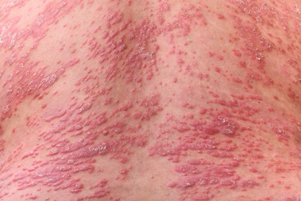
In a recently published paper, researchers provided a review of current knowledge regarding IL-29. Read what they found below. (©Sweetheart/StudioShutterstock.com)

Interleukin-29 (IL-29) is a newly discovered cytokine that has become the subject of increased research attention for its role in inflammation and inflammatory autoimmune disorders and therefore as a target for novel therapeutic development.
In a recently published paper, researchers provided a review of current knowledge regarding IL-29, including information about its binding, signaling, expression, biologic functions, association with inflammatory autoimmune disorders and therapeutic potential. In presenting the information, the authors stated that they hoped it will contribute to future research on IL-29, provide some clues regarding the role of IL-29 in inflammatory autoimmune diseases and give implications for clinical treatment.
RELATED: Psoriasis research advances treatments for inflammatory skin diseases
AN IL-10 CYTOKINE
IL-29 is a type III interferon and belongs to the IL-10 family, which includes a total of nine cytokines. Like all type III interferons, IL-29 mediates signal transduction by binding to its receptors, IL-28R1 and IL-10R2, that are expressed in dendritic cells, T cells, intestinal epithelial cells, leukemia cells, and melanoma cells. Binding of IL-29 activates downstream signaling pathways, including via the JAK-STAT pathway, that leads to the production of a number of inflammatory cytokines and chemokines and also has been shown to result in apoptosis of melanoma cells.
In addition to its antitumor activity, studies indicate that IL-29 has antiviral and immunoregulatory activity, including roles in innate and adaptive immunity. For example, IL-29 stimulation has been shown to enhance toll-like receptor (TLR) 7/8-mediated B cell proliferation, induce immunoglobulin G and immunoglobulin M production by B cells, and induce IL-10 signaling events in macrophages. It also regulates the production of inflammatory cytokines from monocytes, plasmacytoid dendritic cells, neutrophils, and T cells, and inhibits T cell differentiation. In addition, IL-29 is released by mast cells, induces mast cell infiltration, and regulates the release of various cytokines from mast cells.
The antiviral activity of IL-29 has been shown in studies of its effects on keratinocytes. IL-29 induced expression of TLR3 in keratinocytes as well as expression of interferon-β induced by herpes simplex virus type 1, protecting the keratinocytes from viral attack.
INFLAMMATORY AUTOIMMUNE DISEASES
Expression of IL-29 has been shown to be dysregulated in atopic dermatitis, psoriasis, and other inflammatory autoimmune diseases, including rheumatoid arthritis, systemic lupus erythematosus, osteoarthritis, Sjögren’s syndrome, and systemic sclerosis. In patients with psoriasis, IL-29 expression was found to be upregulated in lesional skin, and the levels of IL-29 expression correlated with antiviral protein expression. Compared with healthy controls, patients with psoriasis were also found to have higher serum levels of IL-29. In addition, IL-29 expression was shown to be increased in the skin of patients with atopic dermatitis compared with healthy controls.
Studies of patients with rheumatoid arthritis, systemic lupus erythematosus, osteoarthritis, Sjogren’s syndrome, Hashimoto’s thyroiditis, and systemic sclerosis also show increased expression of IL-29 in the serum, whereas a decreased level of IL-29 expression was seen in ocular fluid of patients with juvenile idiopathic arthritis-associated uveitis compared with ocular fluid from controls with idiopathic uveitis.
RELATED: Atopic dermatitis treatment advances on psoriasis research
In an animal model, injection of IL-29 into the skin induced local expression of C-X-X motif chemokines 10 and 11 and T cell infiltration, leading to skin swelling. In psoriatic lesional skin, neutralization of IL-29 with an anti-IL-29 antibody resulted in downregulation of antiviral protein expression.
Based on the accumulating evidence that indicates IL-29 has a role in the pathogenesis of inflammatory autoimmune disorders, researchers suggest that the development of the autoimmune diseases might be suppressed by interventions that reverse abnormal IL-29 expression. Recognizing, however, that the pathogenesis of inflammatory autoimmune diseases is complex, the authors noted that further studies are needed to understand the role and therapeutic effects of IL-29.
References:
Wang JM, Huang AF, Xu WD, Su LC. Insights into IL-29: Emerging role in inflammatory autoimmune diseases. J Cell Mol Med. 2019;


